Stability of Self-Assembled Organic-Inorganic Layered Perovskite
- PDF / 753,637 Bytes
- 6 Pages / 417.6 x 639 pts Page_size
- 48 Downloads / 218 Views
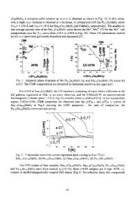
EXPERIMENTAL PROCEDURE Thin films of microcrystalline PhE-PbX4 (X; I and Br, about 20 nm in thickness) were fabricated on a Si0 2 glass substrate by the spin-coating method. The 2 wt% NNdimethylformamide (DMF) solutions, dissolved stoichiometric amounts of C6H 5 C2H4NH 3X and PbX 2, were used in this study. Using these solutions, spin-coating was carried out at 4000 rpm for 15 sec. The substrate surface temperature was kept at 60 °C during spinning. The photostability of the samples was estimated from the relative exciton absorption intensity (I/Io) as a function of photo-irradiation time. Note that Io means an exciton absorption intensity of as-prepared film and I, means that of irradiated for t mrin. Exciton absorption intensity of the samples was measured using a conventional Vis-UV spectrophotometer at room temperature. The samples were irradiated by the light beam which was monocromated using a band-pass filter (U-34) and focused to 1 mm in diameter. High pressure mercury lamp (500 W) was used as a UV-irradiation source. The photo-irradiation was carried out in an atmospheric pressure and vacuum (approximately 0.1 Pa) at room temperature. The crystallinity and orientation of the samples before and after irradiation were characterized by X-ray diffraction (XRD, Rigaku, RINT2500 X-ray diffractometer) using a monochromated CuKat radiation (30 kV, 20 mA). The near-surface composition of the samples was estimated by using X-ray photoelectron spectroscopy (XPS, PHI Model-1700) using a non-monochromated MgKct radiation (15 kV, 26 mA). RESULTS AND DISCUSSION Photo-stability X-ray diffraction patterns of PhE-PbX 4 films revealed that these samples fabricated by the spin-coating method were single-phase and highly oriented, with the c-axis perpendicular to the substrate surface. Figure 1 shows the Vis-UV absorption spectra of PhE-PbI4 films irradiated in (a) air and (b) in vacuum for various periods. As seen in the Fig l(a), the as-prepared film showed strong absorption band with the narrow bandwidth at 518 nm, which have been attributed to the exciton formed in the [Pb614 layer by the transition from Pb 2'(6s) to Pb 2 +(6p) orbital [4]. With increasing photo-irradiation time, the exciton absorption band decreased rapidly and no typical absorption band was observed after irradiation. In Fig l(b), decrease in the exciton absorption band was also observed, but, a new absorption band appeared at around 410 nm with increasing irradiation time. The spectra considerably resemble as aniline (C6H5 NH 2 ) intercalated PbI 2 [9].
166
0.5
0.5
0.4
0.4
0.3
0.3
0.2
0.2
() C
(U .0 M 0
U)
20 Mmn,•j
0.1
400
500
600
700
800
400
Wavelength (nm)
500
S0.1 600
700
800
Wavelength (nm)
Fig. 1. Vis-UV absorption spectra of PhE-PbI 4 films irradiated in (a) air and (b) vacuum for various periods.
Changes
in
the
relative
exciton
absorption intensity (II) of the PhE-PbX 4 (X; Br, I) films as a function of photo-irradiation time are shown in Fig 2. In the PhE-Pb14 films, the I/A. rapidly decreased with the photo-ir
Data Loading...