T2-weighted cardiovascular magnetic resonance in acute cardiac disease
- PDF / 2,445,692 Bytes
- 11 Pages / 595.276 x 793.701 pts Page_size
- 26 Downloads / 325 Views
REVIEW
Open Access
T2-weighted cardiovascular magnetic resonance in acute cardiac disease Ingo Eitel1,2, Matthias G Friedrich2*
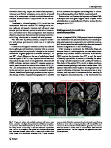
Abstract Cardiovascular magnetic resonance (CMR) using T2-weighted sequences can visualize myocardial edema. When compared to previous protocols, newer pulse sequences with substantially improved image quality have increased its clinical utility. The assessment of myocardial edema provides useful incremental diagnostic and prognostic information in a variety of clinical settings associated with acute myocardial injury. In patients with acute chest pain, T2-weighted CMR is able to identify acute or recent myocardial ischemic injury and has been employed to distinguish acute coronary syndrome (ACS) from non-ACS as well as acute from chronic myocardial infarction. T2-weighted CMR can also be used to determine the area at risk in reperfused and non-reperfused infarction. When combined with contrast-enhanced imaging, the salvaged area and thus the success of early coronary revascularization can be quantified. Strong evidence for the prognostic value of myocardial salvage has enabled its use as a primary endpoint in clinical trials. The present article reviews the current evidence and clinical applications for T2-weighted CMR in acute cardiac disease and gives an outlook on future developments. “The principle of all things is water” Thales of Miletus (624 BC - 546 BC) Introduction Cardiovascular magnetic resonance (CMR) is wellestablished and increasingly used in clinical practice for the diagnosis and management of cardiovascular disease [1-3]. Importantly, recent technological advances of CMR have introduced its use for visualizing certain tissue changes in patients with acute myocardial diseases. This is of particular interest in patients with suspected ischemic disease, a broad and heterogeneous population that challenges the clinician in terms of: 1) accurately establishing the diagnosis; 2) risk stratification; 3) therapeutic decision making; and 4) monitoring response to therapy [4]. CMR is uniquely able to integrate, in a single examination, an accurate quantitative assessment of left ventricular (LV) function, structural abnormalities of the myocardial tissue including edema, infarct size, and myocardial salvage as well as its microvascular status. * Correspondence: [email protected] 2 Stephenson Cardiovascular Magnetic Resonance Centre at the Libin Cardiovascular Institute of Alberta, Departments of Cardiac Sciences and Radiology, University of Calgary, Calgary, Canada Full list of author information is available at the end of the article
Therefore, CMR has an unparalleled potential as the main diagnostic tool in acute cardiac disease by providing information on the stage, degree, and extent of reversible and irreversible myocardial injury [5,6]. Specifically, T2-weighted CMR has recently generated significant interest and has been employed to distinguish acute coronary syndrome (ACS) from non-ACS and recent from remote infarction in patients with undifferentiated ches
Data Loading...