Temperature and Orientation Effects in Iron Nitride Crystal Formation
- PDF / 212,219 Bytes
- 5 Pages / 420.48 x 639 pts Page_size
- 111 Downloads / 376 Views
TEMPERATURE AND ORIENTATION EFFECTS IN IRON NITRIDE CRYSTAL FORMATION
L. J. LOWDER, W. FRANZEN, AND R. J. CULBERTSON Army Materials Technology Laboratory, Watertown, MA 02172-0001
ABSTRACT Pure rolled iron foils 0.025 mm thick have been implanted at two different angles of incidence and at several different temperatures. The implantation dose in each case was 5x1 017 atoms/cm 2 . Both implanted and unimplanted foils were analyzed by transmission x-ray diffraction. Foils implanted at 320 ± 5°C exhibit peaks that correspond to the formation of several different phases of iron-nitride crystals, as observed by other investigators.[ 1,2] No such formation takes place at an implantation temperature of-20'C. We have evidence that the orientation of the iron nitride crystals is correlated with the orientation of the iron crystals.
INTRODUCTION The morphology of micro-crystals of iron nitride formed during nitrogen implantation of iron is interesting because of its relation to the change in mechanical properties (hardness, friction, wear) brought about by the implantation. Temperature is an important parameter in this connection because the sample may be heated by the ion beam during implantation or in later use by mechanical working. Rauschenbach and his collaborators [ 1,3-7] have carried out an extensive series of studies on the crystal structure of nitrogen-implanted iron as a function of temperature and other parameters. From their work and subsequent work carried out in this laboratory [2] it is known that the growth of the metastable phase y '-Fe 4 N tends to be favored at implantation temperatures above 300'C. Since the first-order maximum that corresponds to diffraction from the (200) planes of y '-Fe 4N can be clearly resolved from other structures in the diffraction pattern, we decided to investigate the morphology of micro-crystals of this substance formed in iron when implanted with nitrogen at different temperatures, using a transmission x-ray spectrometer as an analytical tool. Other phases of iron nitride are not as well resolved from one another, nor from the carbo-nitrides that have been reported by some investigators [3].
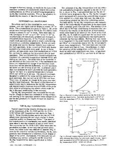
EXPERIMENT The implanted iron was in the form of 0.025 mm thick high-purity Johnson Matthey Foils. The total impurity content of these foils does not exceed 30 ppm, with no single impurity element present at a concentration greater than 5 ppm. Foils were mounted on a temperature-controlled sample holder consisting of a shielded copper plate 1.2 cm thick that could be heated by a 4 kV electron beam from the rear, or cooled by thermal links to a cold reservoir, as shown in Fig. 1. The implanter used is a Zymet Z- 100 machine that generated an unanalyzed beam consisting of 60% N2 ' and 40% N' ions at 80 keV. The x-ray diffractometer used is a Picker diffractometer set up in transmission using Mo Ka radiation (X=0.7093A). A feature of the spectrometer is that it allows the orientation of the crystallites formed during implantation to be investigated by changing the foil-tilt angle w (see
Data Loading...