Temporary exacerbation of benign external hydrocephalus following minor head trauma
- PDF / 374,426 Bytes
- 2 Pages / 595.276 x 790.866 pts Page_size
- 61 Downloads / 291 Views
LETTER TO THE EDITOR
Temporary exacerbation of benign external hydrocephalus following minor head trauma Keyvan Tayebi Meybodi 1 & Zohreh Habibi 1
&
Farideh Nejat 1
Received: 19 June 2020 / Accepted: 24 June 2020 # Springer-Verlag GmbH Germany, part of Springer Nature 2020
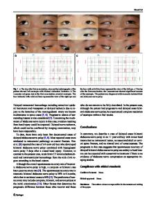
Dear Editor: We would like to share our clinical experience in a subset of infants with incidentally found external hydrocephalus who developed signs and symptoms of raised intracranial pressure (RICP) within a few days after a minor head trauma for a short while. An infant was admitted to the emergency department of our hospital with a minor head trauma. On examination, the infant was alert. The head was slightly large for age according to the standard curves. Fontanelle was relaxed. No neurologic deficit was present. On head computed tomography (CT), a small rim of extra-axial hematoma was evident along with a wide subarachnoid space in the frontal and anterior interhemispheric region, consistent with radiologic external hydrocephalus (EH) (Fig. 1a). No intervention except close clinical monitoring was offered. After a few days, the patient was readmitted with repeated emesis, no tolerance to oral feeding, and a full, tense fontanelle. A repeated head CT revealed no new finding except exacerbated widening of subarachnoid space (Fig. 1b). After a subdural tap for acutely increased intracranial hypertension, conservative therapy including intravenous hydration and antiemetic drug therapy was successful in controlling the emesis. With oral tolerance, acetazolamide was prescribed on discharge. Close follow-up during 6 months after discharge disclosed relaxed fontanelle with normal rate of head growth. No further CT scan was requested since the patient remained symptom free. We have encountered multiple large-headed infants with undiagnosed asymptomatic EH who, after a minor head trauma, presented with sign and symptoms of RICP or soaring growth rate of head circumference. None of them had previous history of neurological dysfunction. On head CT, all of * Zohreh Habibi [email protected] 1
Department of Neurosurgery, Children’s Hospital Medical Center, Tehran University of Medical Sciences, No. 62, Qarib St., Keshavarz Blvd., Tehran 1419733151, Iran
them had frontal expansion of subarachnoid space, suggestive of benign idiopathic EH. All of them were managed with conservative therapy with diuretics. No one required a subduroperitoneal shunt. Increased HC may be caused by three different mechanisms: increased skull thickness, enlargement of cerebrospinal fluid (CSF) spaces, or increased brain parenchyma. Intracranial CSF spaces are subdivided to ventricles and subarachnoid space (SAS). External hydrocephalus (EH) is defined as increased HC predominantly due to expansion of intracranial SAS, more at frontal lobe and anterior interhemispheric fissure, with or without mild ventriculomegaly. There are some controversies pertaining to the definition, as some authors insist upon the value of symptoms and signs of raised intracranial pressure (
Data Loading...