The precision of patient-specific instrumentation guides for the positioning of the glenoid component in total reverse s
- PDF / 524,818 Bytes
- 6 Pages / 595.276 x 790.866 pts Page_size
- 101 Downloads / 262 Views
ORIGINAL PAPER
The precision of patient-specific instrumentation guides for the positioning of the glenoid component in total reverse shoulder arthroplasty: an in vivo scanographic study Axel Marcoin 1 & Cécile Nerot 1 & Thibault Lestra 1 & Laurent Blasco 1 & Antoine Ferrier 1 & Renaud Siboni 1 & Xavier Ohl 1 Received: 11 October 2019 / Accepted: 4 March 2020 # SICOT aisbl 2020
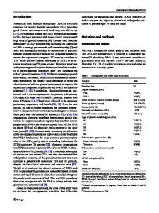
Abstract Objective Optimal position and fixation of the baseplate is essential for the longevity of the reverse shoulder arthroplasty (RSA) and the patient-specific instrumentation (PSI) can help the surgeon to achieve that purpose. The aim of this study was to assess the reliability of the PSI guides for the positioning of the baseplate and the fixation’s screws. Method Prospective study involving 35 patients operated for RSA. The PSI guides were planned and used by the senior surgeon in all cases. We compared the planned orientation (frontal and axial) of the baseplate and the screws with the post-operative CT scan. Results The mean difference between the planned measures and the post-op measures was inferior to 2.5°. The screw’s length corresponded with the pre-op plan in 70% of the cases. Conclusion The use of a PSI guide to position the glenoid implant in total reverse shoulder arthroplasty is reliable, reduces the risk of positioning errors and improves the quality of fixation with the screws. Keywords Reverse shoulder arthroplasty . 3D planification . Patient-Specific Instrumentation (PSI) . Glenoid orientation . CT-Scan
Introduction Total reverse shoulder arthroplasty (TSA) is recognized as the choice treatment for cuff tear arthropathy or massive cuff tear [1–3]. The occurrence of scapular notching is frequent and potentially leads to glenoid migration. This complication is essentially due to mechanical impingement between the medial part of the implant and the scapular pillar in adduction position [4]. Numerous authors have suggested modifications to the surgical technique to avoid this stress, in particular, via an improvement in the positioning of the baseplate without affecting the quality of the fixation [4–6]. * Xavier Ohl [email protected] 1
Hopital Maison-Blanche, CHU de Reims, 45 rue Cognacq-Jay, REIMS Cedex, 51092 Reims, France
Pre-operative 2D or even 3D planning is a first strategy. Peri-operative navigation is a reliable option, but it requires the positioning of optical markers and the use of specialized equipment in the operating theatre, as well as a learning curve. Finally, several manufacturers offer specific instrumentation (patient-specific instrumentation, PSI) designed and manufactured pre-operatively from a CT scan. The first in vitro studies have shown good reliability for these specific PSI guides [7–9]. Cadaveric and in vivo studies [7–11] have evaluated and compared the quality of the positioning of the baseplate without PSI (standard technique) and with PSI. None of these studies has assessed the positioning of the screws (for length and orientation). Our hypothesis was that the PSI technique would e
Data Loading...