Uncharted territories: assessing cerebral vascularization in patients undergoing venoarterial extra-corporeal membrane o
- PDF / 837,676 Bytes
- 2 Pages / 595.276 x 790.866 pts Page_size
- 76 Downloads / 263 Views
IMAGING IN INTENSIVE CARE MEDICINE
Uncharted territories: assessing cerebral vascularization in patients undergoing venoarterial extra‑corporeal membrane oxygenation Paul‑Henri Wicky1, Marie‑Cécile Henry‑Feugeas2, Marylou Para3 and Etienne de Montmollin1,4* © 2020 Springer-Verlag GmbH Germany, part of Springer Nature
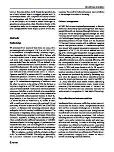
A 39-year-old woman was placed under femoro-axillary venoarterial extra-corporeal membrane oxygenation (VA-ECMO) (tip positions: right subclavian artery/inferior vena cava, subhepatic veins level) for graft dysfunction after cardiac transplantation. Eleven days later, a cervicocephalic computed tomography angiography (CTA) with contrast injection through a left subclavian central venous catheter was performed, while VA-ECMO flow rate was 3.1 L/min. There was no opacification of the right internal carotid and right middle cerebral artery (MCA) (Fig. 1a, c), despite present blood flow assessed by Doppler ultrasonography in internal carotids and MCA. The patient did not present any neurological deficit. Due to improving cardiac function, VA-ECMO was removed
*Correspondence: [email protected] 1 Medical and Infectious Diseases ICU, Bichat‑Claude Bernard Hospital, Assistance Publique-Hôpitaux de Paris, Paris, France Full author information is available at the end of the article
24 h later. A subsequent CTA showed normal opacification of the right carotid territory, confirming the initial misestimation of cerebral perfusion (Fig. 1b, d). Lower ECMO pump flow and intrinsic cardiac output resulted in differential opacifications of the carotid arteries: right-sided perfusion from ECMO-injected, non-contrasted blood originating from the inferior vena cava and left-sided cardiac-contrasted blood from the superior vena cava. These phenomena depend on arterial and venous cannula location, ECMO pump flow and site of contrast bolus injection. Intensivists and radiologists should be aware of these potential pitfalls of CTA head imaging in VA-ECMO patients.
Fig. 1 a and b Axial plane cephalic computed tomography angiography before and after VA-ECMO removal. Dashed blue arrow: absence of opacification of the right middle cerebral artery. Plain blue arrow: re-opacified right middle cerebral artery after VA-ECMO removal. c and d Coronal plane cervicocephalic computed tomography angiography before and after VA-ECMO removal. Red arrow: right axillary artery VA-ECMO reinjection cannula. Dashed orange arrow: absence of opacification of the right internal carotid artery. Plain orange arrow: reopacified right internal carotid artery after VA-ECMO removal.
Author details 1 Medical and Infectious Diseases ICU, Bichat‑Claude Bernard Hospital, Assistance Publique-Hôpitaux de Paris, Paris, France. 2 Department of Radiol‑ ogy, Bichat‑Claude Bernard Hospital, Assistance Publique-Hôpitaux de Paris, Paris, France. 3 Department of Cardiothoracic Surgery, Bichat‑Claude Bernard Hospital, Assistance Publique-Hôpitaux de Paris, Paris, France. 4 UMR 1137, IAME Team5‑DeScID, Université́ de Paris, Paris, France. Compliance
Data Loading...