Unmasking Of pathologic Q waves by left bundle branch pacing
- PDF / 708,117 Bytes
- 2 Pages / 595.276 x 790.866 pts Page_size
- 69 Downloads / 264 Views
CASE REPORTS
Unmasking Of pathologic Q waves by left bundle branch pacing Shunmuga Sundaram Ponnusamy 1
&
Pugazhendhi Vijayaraman 2
Received: 17 August 2020 / Accepted: 4 September 2020 # Springer Science+Business Media, LLC, part of Springer Nature 2020
A 61-year-old man with left ventricular ejection fraction of 25% was referred for cardiac resynchronization therapy. He had undergone percutaneous coronary intervention to right coronary artery (RCA) for inferior wall myocardial infarction 3 years ago. Electrocardiography (ECG) showed complete left bundle branch block with QRS duration of 174 ms (Fig. 1a). Coronary angiography confirmed patent stent in RCA. Left bundle branch pacing (LBBP) was done using C315 sheath and 3830 Selectsecure™ lead (Medtronic, Minneapolis, MN) [1] (Fig. 2a). Nonselective to selective capture of left bundle could be demonstrated at near pacing threshold value. Paced
* Shunmuga Sundaram Ponnusamy [email protected] 1
Department of Cardiology, Velammal Medical College Hospital and Research Institute, Madurai, Tamil Nadu, India
2
Geisinger Commonwealth School of Medicine, Geisinger Heart Institute, MC 36-10, 1000 E Mountain Blvd, Wilkes-Barre, PA 18711, USA
ECG showed rSR pattern in lead V1 with QRS duration of 108 ms (Fig. 2b) and peak left ventricular activation time of 65 ms. Inferior leads showed resurgence of Q wave (> 40 ms) corresponding to the old inferior wall myocardial infarction(Fig. 1b and 2b). ECG diagnosis of myocardial ischemia during right ventricular pacing and native left bundle branch block can be difficult as non-physiological activation would result in masking of pathological Q waves and ST segment changes [2]. LBBP restores the physiological activation of the ventricle thereby unmasking the pathological Q waves.
J Interv Card Electrophysiol
Fig. 1 a Baseline 12 lead ECG showing complete LBBB with QRS duration of 174 ms without Q wave in inferior lead. b Post-LBBP paced ECG showing narrow QRS (duration 102 ms) with pathological
Q in inferior leads and T-wave memory. Right bundle branch delay due to LBBP was corrected by optimizing the AV delay
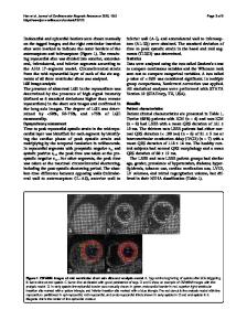
Fig. 2 a Fluoroscopic view in left anterior oblique view showing the depth of 3830 pacing lead inside the proximal septum. b Unipolar paced ECG showing rSR in lead V1 with rapid left ventricular activation time (65 ms). Note the distinct isoelectric interval between
pacing spike and onset of QRS complex. Pathological Q wave (> 40ms duration) noted in inferior leads. HB, his bundle; RVA, right ventricular apex; LBBP, pacing lead
References Compliance with ethical standards 1. The study was conducted after getting the ethical committee approval. The paper is not under consideration elsewhere. None of the paper’s contents has been previously published. All authors have read and approved the manuscript. Conflict of interest The authors declare that they have no conflict of interest.
2.
Ponnusamy SS, Arora V, Namboodiri N, Kumar V, Kapoor A, Vijayaraman P. Left bundle branch pacing: a comprehensive review. J Car
Data Loading...