Use of Soft Lithography for Multi-layer MicroMolding (MMM) of 3-D PCL Scaffolds for Tissue Engineering
- PDF / 1,170,888 Bytes
- 6 Pages / 612 x 792 pts (letter) Page_size
- 63 Downloads / 262 Views
W9.3.1/O5.3.1
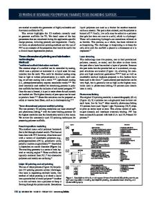
Use of Soft Lithography for Multi-layer MicroMolding (MMM) of 3-D PCL Scaffolds for Tissue Engineering Yang Sun, Nicholas Ferrell, and Derek J. Hansford Biomedical Engineering Center, The Ohio State University, 270 Bevis Hall, 1080 Carmack Rd., Columbus, OH 43210, U.S.A. ABSTRACT Tissue engineering scaffolds with precisely controlled geometries, particularly with surface features smaller than typical cell dimensions (1-10µm), can improve cellular adhesion and functionality. In this paper, soft lithography was used to fabricate polydimethylsiloxane (PDMS) stamps of arrays of parallel 5µm wide, 5µm deep grooves separated by 45 µm ridges, and an orthogonal grid of lines with the same geometry. Several methods were compared for the fabrication of 3-D multi-layer polycaprolactone (PCL) scaffolds with precise features. First, micromolding in capillaries (MIMIC) was used to deliver the polymer into the small grooves by capillarity; however the resultant lines were discontinuous and not able to form complete lines. Second, spin coating and oxygen plasma were combined to build 3-D scaffolds with the line pattern. The resultant scaffolds had good alignment and adhesion between layers; however, the upper layer collapsed due to the poor mechanical rigidity. Finally, a new multi-layer micromolding (MMM) method was developed and successfully applied with the grid pattern to fabricate 3-D scaffolds. Scanning electron microscopy (SEM) characterization showed that the multi-layered scaffolds had high porosity and precisely controlled 3-D structures.
INTRODUCTION Tissue engineering is a practical and promising approach to address the problems of scarcity of donor organs for allograft treatment [1]. Scaffolds with 3-D structures have been applied to support cell growth in wound healing and tissue regeneration [2,3]. In addition, scaffolds have been shown to have effects on cell behaviors, such as proliferation, migration and differentiation [4-6]. Research on contact guidance has shown that line patterns with the feature range of 1-50 microns accelerate cell motion and orient cell location [7-9]. These effects are greatly enhanced when cells grow on precise features, which modulate cytoskeletal structures to control cellular morphology. Since the 1960’s, photolithography and microfabrication have been demonstrated as effective techniques to fabricate surface features in micro- or nano-scales [1012]. It has been demonstrated that cell growth, migration, proliferation, and differentiation are regulated by the surface topographical factors such as ridges, islands, wells, etc. [13-15]. For the practical applications of tissue engineering, it is highly desirable to fabricate 3-D scaffolds with designed geometrical properties to elicit appropriate cell behavior. However, the precise control of microfeature geometry remains a difficult problem [16,17]. Three-dimensional scaffolds with feature sizes of 50-100µm, or even several hundreds of microns have limits when studying individual cell behaviors and are not capable of preci
Data Loading...