Wrist and Hand
The wrist and hand is an excellent region for the use of musculoskeletal ultrasound due to the superficial structures and the ability to perform dynamic scans. Common pathologies in this area include tenosynovitis of the dorsal wrist compartments, tendon
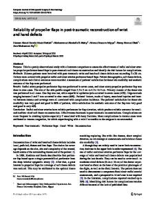
- PDF / 2,210,098 Bytes
- 13 Pages / 595.28 x 790.87 pts Page_size
- 25 Downloads / 318 Views
Wrist and Hand David A. Spinner and Melissa I. Rosado
The wrist and hand is an excellent region for the use of musculoskeletal ultrasound due to the superficial structures and the ability to perform dynamic scans. Common pathologies in this area include tenosynovitis of the dorsal wrist compartments, tendon ruptures, cysts, compression neuropathies, and arthritides.
Carpal Tunnel Syndrome The carpal tunnel is located on the volar aspect of the wrist and is the most common compression neuropathy site in the upper limb [1]. Carpal tunnel syndrome (CTS) is a constellation of symptoms consisting of pain, paresthesias, and eventually thenar atrophy, arising from increased pressure within the tunnel, edematous states, or direct nerve trauma. Patients often complain of nocturnal paresthesias in the volar aspect of the thumb, index finger, middle finger, and radial half of the ring finger. Provocative tests such as compression over the carpal tunnel or Tinel’s and Phalen’s signs can help to reproduce the symptoms. The diagnosis is based on history, physical exam, electrodiagnostic studies, or cross-sectional area on ultrasound [2]. Corticosteroid injection into the carpal tunnel has been shown to provide improvement in pain, paresthesias, and function [3–5].
Scanning Technique and Anatomy to Identify The patient should sit with the elbow flexed to 90°, forearm supinated with the hand resting comfortably. A towel may be placed under the wrist to place it in slight extension. The carpal tunnel lies just distal to the distal wrist crease. The transducer
D.A. Spinner, DO, RMSK (*) Department of Anesthesiology – Pain Medicine, Arnold Pain Management Center, Beth Israel Deaconess Medical Center, Harvard Medical School, Brookline, MA, USA e-mail: [email protected] M.I. Rosado, MD Maxwell Medical, New York, NY, USA
is placed transversely (short axis) to the median nerve, at the distal wrist crease. The bony borders of the tunnel include the scaphoid and trapezium laterally and the hamate and pisiform medially. The transverse carpal ligament or flexor retinaculum forms the superficial roof of the tunnel. The carpal tunnel contains the tendons of the flexor digitorum profundus, flexor digitorum superficialis, and flexor pollicis longus, and the median nerve. Identify the honeycomb-appearing median nerve in cross section. It generally appears relatively hypoechoic to the adjacent hyperechoic tendons in cross section. You can tilt the probe to adjust anisotropy; the nerve will remain present, but the flexor tendons may disappear at off angles. Have the patient move their flexor tendons to assess for adhesions which may be amenable to hydrodissection. Be sure to scan on the ulnar side of the canal to view the ulnar artery and nerve and radially to identify the radial artery (Fig. 4.1) [6, 7].
Injection Techniques: In-Plane Axial UlnarSided Approach [8] Patient positioning: Sit the patient with the affected arm resting comfortably on the table. A towel can be placed underneath the wrist to create mild extension. Probe position:
Data Loading...