1039 Prevalence of cardiovascular manifestations in patients with Marfan syndrome: a cardiovascular magnetic resonance s
- PDF / 98,184 Bytes
- 2 Pages / 610 x 792 pts Page_size
- 35 Downloads / 377 Views
BioMed Central
Open Access
Meeting abstract
1039 Prevalence of cardiovascular manifestations in patients with Marfan syndrome: a cardiovascular magnetic resonance study Francisco Alpendurada* and Raad Mohiaddin Address: Royal Brompton Hospital, London, UK * Corresponding author
from 11th Annual SCMR Scientific Sessions Los Angeles, CA, USA. 1–3 February 2008 Published: 22 October 2008 Journal of Cardiovascular Magnetic Resonance 2008, 10(Suppl 1):A164
doi:10.1186/1532-429X-10-S1-A164
Abstracts of the 11th Annual SCMR Scientific Sessions - 2008
Meeting abstracts – A single PDF containing all abstracts in this Supplement is available here. http://www.biomedcentral.com/content/pdf/1532-429X-10-S1-info.pdfThis abstract is available from: http://jcmr-online.com/content/10/S1/A164 © 2008 Alpendurada and Mohiaddin; licensee BioMed Central Ltd.
Introduction Marfan syndrome (MS) is a rare connective tissue disease (incidence 1/10,000 persons) caused by mutations of the fibrillin-1 gene. Although it can affect several systems, the cardiovascular system is the major source of morbidity and mortality. As a result, Cardiovascular Magnetic Resonance (CMR) can play an important role in identifying and evaluating cardiovascular manifestations in this population.
Methods The study population consisted of 120 consecutive Marfan patients referred to our centre between January 2003 and June 2007 including 77 males (64%) and 43 females (36%), with a mean age of 34.9 years. We evaluated thoracic aortic dimensions at different segments, and assessed for structural abnormalities of the aorta, aortic arch branches, and main pulmonary artery. Aortic and mitral valves anatomy and function were also evaluated. In one study both valves were not evaluated due to incomplete study caused by claustrophobia, while in another study the mitral valve was not assessed due to technical artifact. Although it was not the main objective of this study, assessment of chest wall deformities and pulmonary abnormalities was also performed.
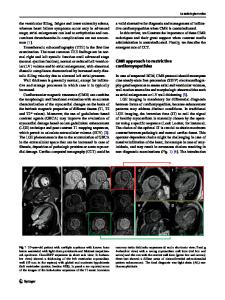
patients (19.2%) average diameter 32.7 cm, the aortic arch was dilated in 19 patients (15.8%), average diameter 23.7 cm, and the descending aorta was dilated in 18 patients (15.0%), average diameter 23.8 cm. The arch vessels were dilated or aneurysmatic in 9 patients (7.5%), and the abdominal aorta was involved in 9 patients (7.5%). The main pulmonary artery was dilated in 16 patients (13.3%). Aortic dissection was noted in 9 patients (7.5%): one acute type A dissection, 7 chronic type A dissections, and 1 chronic type B dissection. Intramural haematomas were seen in 2 patients. There was aortic regurgitation in 50 out of the 101 patients with native aortic valve (49.5%), which was moderate or severe in 14 patients (13.9%). The native aortic valve was bicuspid in 4 patients. Of the 116 patients with identifiable native mitral valve, 43 patients (37.1%) met criteria for mitral valve prolapse. Mitral regurgitation was identified in 22 patients (19.0%), which was moderate or severe in 8 patients (6.9%). Fifty
Data Loading...