3D construct of hydroxyapatite/zinc oxide/palladium nanocomposite scaffold for bone tissue engineering
- PDF / 4,083,547 Bytes
- 14 Pages / 595.276 x 790.866 pts Page_size
- 78 Downloads / 361 Views
TISSUE ENGINEERING CONSTRUCTS AND CELL SUBSTRATES Original Research
3D construct of hydroxyapatite/zinc oxide/palladium nanocomposite scaffold for bone tissue engineering Fatemeh Heidari 1 Fahimeh Sadat Tabatabaei2,3 Mehdi Razavi4 Reza Bazargan Lari5 Mina Tavangar1 Georgios E. Romanos6 Daryoosh Vashaee7 Lobat Tayebi3 ●
●
●
●
●
●
●
1234567890();,:
1234567890();,:
Received: 16 February 2020 / Accepted: 12 July 2020 © Springer Science+Business Media, LLC, part of Springer Nature 2020
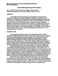
Abstract The purpose of this study was to produce and characterize Hydroxyapatite/Zinc Oxide/Palladium (HA/0.05 wt% ZnO/0.1 wt % Pd) nanocomposite scaffolds and study their mechanical and antibacterial properties, biocompatibility and bioactivity. The initial materials were developed using sol-gel and precipitation methods. Scaffolds were characterized using atomic absorption analysis (AA), scanning electron microcopy (SEM), energy dispersive spectroscopy (EDS) and transmission electron microscopy (TEM), atomic force microscopy (AFM) and Brunauer−EmmeS−Teller (BET) method. Furthermore, the bioactivity of scaffolds in simulated body fluid (SBF) and the interaction of dental pulp stem cells (DPSCs) with the nanocomposite scaffolds were assessed. Our results showed that the HA/ZnO/Pd (H1), HA/ZnO/Pd coated by 0.125 g chitosan (H2) and HA/ZnO/Pd coated by 0.25 g chitosan (H3) scaffolds possess higher compressive strength and toughness and lower microhardness and density compared to the pure HA (H0) scaffolds. Immersion of samples in SBF showed the deposition of apatite on the surface of the scaffolds. The biocompatibility assay indicated lower cell proliferation on the H1, H2 and H3 in comparison to the H0. The antibacterial results obtained show a significant impact by loading Pd/ZnO on HA in the deactivation of microorganisms in vitro. Graphical Abstract
Compressive strength (MPa)
CS Tsintering= 1300 °C
HA
290
315
HA 22.4 HA
HA/ZNO/PD
HA/ZNO/PD/CS
Pd ZnO
CS/HA/ZnO/Pd
MIC and MBC Assay
* Fatemeh Heidari [email protected] 1
Department of Materials Engineering, School of Engineering, Yasouj University, Yasouj 75918-74934, Iran
2
Department of Dental Biomaterials, School of Dentistry, Shahid Beheshti University of Medical Sciences, Tehran, Iran
3
Marquette University School of Dentistry, Milwaukee, WI 53233, USA
4
Department of Radiology, School of Medicine, Stanford University, Palo Alto, CA 94304, USA
5
Department of Materials Science and Engineering, Marvdasht Branch, Islamic Azad University, Marvdasht, Iran
6
Stony Brook University, School of Dental Medicine, Stony Brook, NY 11794, USA
7
Department of Electrical and Computer Engineering, NC State University, Raleigh, NC 27695, USA
85 Page 2 of 14
Journal of Materials Science: Materials in Medicine (2020)31:85
1 Introduction Tissue engineering offers a novel approach in reproducing new tissues for use in repair and regeneration of damaged organs [1]. This approach shows promising results in treating bone defects and fractures, in which employing hydroxy
Data Loading...