Altered skeletal muscle (mitochondrial) properties in patients with mitochondrial DNA single deletion myopathy
- PDF / 611,886 Bytes
- 12 Pages / 595.276 x 790.866 pts Page_size
- 29 Downloads / 376 Views
RESEARCH
Open Access
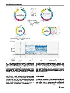
Altered skeletal muscle (mitochondrial) properties in patients with mitochondrial DNA single deletion myopathy Saskia Maria Gehrig1,2,3, Violeta Mihaylova1, Sebastian Frese1, Sandro Manuel Mueller1, Maria Ligon-Auer1, Christina M. Spengler3,4, Jens A. Petersen1, Carsten Lundby2,3 and Hans H. Jung1,3*
Abstract Background: Mitochondrial myopathy severely affects skeletal muscle structure and function resulting in defective oxidative phosphorylation. However, the major pathomechanisms and therewith effective treatment approaches remain elusive. Therefore, the aim of the present study was to investigate disease-related impairments in skeletal muscle properties in patients with mitochondrial myopathy. Accordingly, skeletal muscle biopsies were obtained from six patients with moleculargenetically diagnosed mitochondrial myopathy (one male and five females, 53 ± 9 years) and eight age- and gender-matched healthy controls (two males and six females, 58 ± 14 years) to determine mitochondrial respiratory capacity of complex I-V, mitochondrial volume density and fiber type distribution. Results: Mitochondrial volume density (4.0 ± 0.5 vs. 5.1 ± 0.8 %) as well as respiratory capacity of complex I-V were lower (P < 0.05) in mitochondrial myopathy and associated with a higher (P < 0.001) proportion of type II fibers (65.2 ± 3.6 vs. 44.3 ± 5.9 %). Additionally, mitochondrial volume density and maximal oxidative phosphorylation capacity correlated positively (P < 0.05) to peak oxygen uptake. Conclusion: Mitochondrial myopathy leads to impaired mitochondrial quantity and quality and a shift towards a more glycolytic skeletal muscle phenotype. Keywords: Bioenergetics, Fat oxidation, Mitochondria, Mitochondrial cytopathy, Neuromuscular disease, Skeletal muscle phenotype
Background Despite progress in understanding the biochemistry and genetics of mitochondrial myopathy, many of the pathophysiological mechanisms remain unclear [1]. Validated therapeutic options as well as simple and effective diagnostic tools are lacking [1–5]. So far, it has barely been investigated how muscle metabolism and morphology may be affected by mitochondrial dysfunction. Fiber type abnormalities including varying distribution of type I and II fibers as well as general or selective atrophy have been reported in patients with various mitochondrial respiratory chain dysfunctions [6–8] but not in adults * Correspondence: [email protected] 1 Department of Neurology, University Hospital Zurich, Frauenklinikstrasse 26, 8091 Zurich, Switzerland 3 Zurich Center for Integrative Human Physiology (ZIHP), Winterthurerstrasse 190, 8057 Zurich, Switzerland Full list of author information is available at the end of the article
with mitochondrial myopathy. In a rat model of mitochondrial myopathy [9] a transformation from type I to type II fibers was observed in response to a primary defect of the mitochondrial respiratory chain. In addition, patients with mitochondrial myopathy have been characterised by lower peak oxygen uptake ̇ O2peak) and work
Data Loading...