Amebic Meningoencephalitis Mimicking Tubercular Meningitis
- PDF / 3,532,175 Bytes
- 2 Pages / 595.276 x 790.866 pts Page_size
- 71 Downloads / 309 Views
PICTURE OF THE MONTH
Amebic Meningoencephalitis Mimicking Tubercular Meningitis Sunil Kumar Rao 1
&
Abhishek Abhinay 1 & Pradhap K 1 & Akanksha Singh 1 & Ragini Tilak 2
Received: 26 May 2020 / Accepted: 17 August 2020 # Dr. K C Chaudhuri Foundation 2020
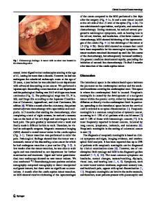
A 16-y-old boy from Bihar presented with intermittent low-grade fever and diffuse headache for 1 mo. The child had a history of one episode of diffuse headache with loss of consciousness for 1 h, 4 mo back and history of swimming in the pond. On examination, the child was conscious; planter extensor, Kernig sign, and neck stiffness were present. Child was managed as a case of septic meningitis with antibiotics as cerebrospinal fluid (CSF) cytology was suggestive of pyogenic picture and no growth was seen in CSF initially and subsequently. But he did not show any improvement in signs and symptoms in 2 wk and repeat CSF cytology showed motile organisms resembling Acanthamoeba in Indian Ink preparation (Fig. 1). Combination of, cotrimoxazole (20 mg/kg/d), rifampicin (10 mg/kg/d) and ketoconazole (5 mg/kg/d) were started but child’s condition was progressively deteriorating. MRI brain showed moderate obstructive hydrocephalus, periventricular ooze, ring enhancing lesion, basal meningeal thickening and enhancement with central transtentorial herniation (Fig. 2). Ventriculo-peritoneal (VP) shunt placement was done and his condition improved. At 4 mo follow-up, the child is doing well without cognitive and neurological deficit with intermittent episodes of headache; CECT brain showed decompressed ventricles and ring enhancing lesion (Fig. 3). Primary amebic meningoencephalitis (PAM) and granulomatous amebic encephalitis (GAE) are rare and fatal clinical entities caused by free-living amebic species in
immunocompetent and immunocompromised individuals [1]. The clinical presentation of PAM and GAE is similar to other central nervous system infections except for their insidious onset, being progressive, not responding to antibiotics, fatal, and to have been identified postmortem. To date no clinical features or CSF findings are characteristic or specific for PAM and GAE, however, intralesional hemorrhage is a recognized imaging and macroscopic finding of GAE [2]. Etiology of acanthameba, in the present case was made by accidental demonstration of trophozoites of acanthameba in wet mount CSF examination with Indian Ink. MRI brain findings mimiced tubercular meningitis [3]. The neutrophilic pleocytosis was seen in CSF cytology; this might be because of pathogenic bacteria inhabitant in ameba which may complicate the actual picture of meningoencephalitis [4]. Clearance of trophozoites from CSF takes from weeks to months [1, 5]; therefore duration of therapy should be individualized.
* Sunil Kumar Rao [email protected] 1
Department of Pediatric Medicine, Institute of Medical Sciences, Banaras Hindu University, Varanasi Uttar Pradesh India
2
Department of Microbiology Institute of Medical Sciences, Banaras Hindu University, Varanasi Uttar Pradesh India
Fig. 1 Troph
Data Loading...