Assessment of pulmonary veins after atrio-pericardial anastomosis by cardiovascular magnetic resonance
- PDF / 336,275 Bytes
- 6 Pages / 595.28 x 793.7 pts Page_size
- 30 Downloads / 303 Views
RESEARCH
Open Access
Assessment of pulmonary veins after atriopericardial anastomosis by cardiovascular magnetic resonance Steven C Greenway1, Shi-Joon Yoo1,2, Giedrius Baliulis3, Christopher Caldarone3, John Coles3 and Lars Grosse-Wortmann1,2*
Abstract Background: The atrio-pericardial anastomosis (APA) uses a pericardial pouch to create a large communication between the left atrium and the pulmonary venous contributaries in order to avoid direct suturing of the pulmonary veins during the repair of congenital cardiac malformations. Post-operative imaging is routinely performed by echocardiography but Cardiovascular Magnetic Resonance (CMR) offers excellent anatomical imaging and quantitative information about pulmonary blood flow. We sought to compare the diagnostic value of echocardiography and CMR for assessing pulmonary vein anatomy after the APA. Methods: This retrospective study evaluated all consecutive patients between October 1998 and January 2010 after either a primary or secondary APA followed by post-repair CMR. Results: Of 103 patients who had an APA, 31 patients had an analyzable CMR study. The average time to CMR was 24.6 ± 32.5 months post-repair. Echocardiographic findings were confirmed by CMR in 12 patients. There was incomplete imaging by echocardiography in 7 patients and underestimation of pulmonary vein restenosis in 12, when compared to CMR. In total, 19/31 patients (61%) from our cohort had significant stenosis following the APA as assessed by CMR. Our data suggest that at least 18% (19/103) of all patients had significant obstruction postrepair. Conclusions: Echocardiography incompletely imaged or underestimated the severity of obstruction in patients compared with CMR. Pulmonary vein stenosis remains a sizable complication after repair, even using the APA.
Background Pulmonary vein (PV) stenosis carries a poor prognosis due to the difficulty of primary surgical repair and the subsequent risk of recurrent obstruction [1-6]. Pulmonary vein stenosis occurs as a complication in a significant fraction of patients post-repair [7], [8]. An alternative to the “classical” operation for PV stenosis, where the PVs are directly anastomosed to the left atrium (LA), is the so-called “sutureless repair” or atrio-pericardial anastomosis (APA) which creates a large communication between the LA and the PVs using a pericardial pouch while avoiding direct suturing of the * Correspondence: [email protected] 1 Division of Cardiology, The Hospital for Sick Children, University of Toronto, Ontario, M5G 1X8, Canada Full list of author information is available at the end of the article
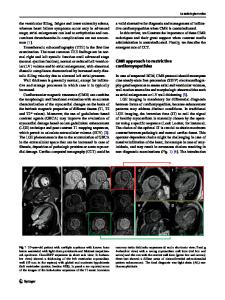
divided edges of the veins [4,5,9-11]. It was hoped that avoiding direct surgical trauma to the veins would reduce the incidence of post-operative obstruction. Since it was first introduced for isolated PV stenosis, the use of the APA has been extended to the correction of partial and total anomalous pulmonary venous connections and short- and medium-term outcomes have been encouraging [4,12-14]. The dominant ima
Data Loading...