Characterization Of Defect Structures in 3C-SiC Single Crystals using Synchrotron White Beam X-Ray Topography
- PDF / 4,008,112 Bytes
- 6 Pages / 414.72 x 648 pts Page_size
- 58 Downloads / 313 Views
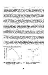
EXPERIMENTAL The crystals analyzed in this work were supplied by the U.S. Army Research Laboratory. They are as-grown, (111) 3C-SiC platelets with sizes of around 3-5 mm across and 1 mm thick. These crystals are transparent and light yellow in color. This color indicates that the crystal is either lightly doped or undoped, since the presence of any metallic impurities or nitrogen introduces a green cast. As the impurity concentration increases the coloration will rapidly approach black [1]. Optical microscopy, in both transmission and reflection modes, shows that these crystals are generally quite uniform, except for occasional inclusions. Scanning Electron Microscope (SEM) observation revealed faceted surfaces, as shown in figure 1. Synchrotron white beam X-ray topography experiments were carried out at the Stony Brook Synchrotron Topography Facility, Beamline X-19C, at the National Synchrotron Light Source (NSLS), Brookhaven National Laboratory. The transmission, or Laue geometry, was employed, with the detector (Kodak SR-5 film) oriented perpendicular to the direction of the area-filling incident beam. Using the synchrotron white beam a single exposure can provide several useful topographic images with linear resolution better than 5 gm. In addition, a new experimental arrangement, involving the use of a narrow beam in reflection geometry, is applied to identify crystal layer structures. As shown schematically in figure 2, the crystal is set parallel to the incident beam and the beam height is adjusted to be smaller than the crystal thickness. The incident beam is controlled to hit only part of the crystal. This way, the diffracted image only shows the structure of the material covered by incident and diffracted beams. Through the precise control of the beam position, a sequence of topographs can be recorded by moving the beam from top to bottom of the crystal. Comparison of these topographs enables one to locate the structural configuration and defect distribution with the crystal. In this study, the beam height is typically 0.2 mm. diffracted beam
crystal thickrness
imaged region incident beam
crystal
beam heightf
diffracted plane Figure. I SEM micrograph of 3C-SiC platelet
Figure 2. Schematic diagram showing the narrow beam reflection geometry.
RESULTS AND DISCUSSION About a dozen crystals were examined. Results from three of them are presented below. Figure 3 shows an X-ray transmission topograph recorded from crystal 1, showing the overall defect distribution. The presence of Pendellosung fringes, labeled F, at the top left edge of the crystal indicates the high quality of this crystal. Stacking faults are found in the middle of the 546
crystal, as indicated by S. Dislocations, indicated by D, are also observed. Partial dislocations bounding the stacking faults are indicated by P and inclusions by I. Some evidence for the existence of growth sector boundaries, G, is also visible. Comparing with figure 1, it is clear that the lines labeled by L in figure 3 are related to the surface step morphology
Data Loading...