Deposition Sequences for Atomic Layer Growth of AlN Thin Films on Si(100) Using Dimethylethylamine Alane and Ammonia
- PDF / 969,530 Bytes
- 6 Pages / 414.72 x 648 pts Page_size
- 44 Downloads / 281 Views
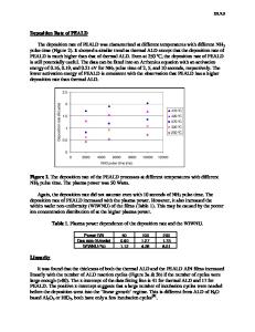
EXPERIMENT Experiments were conducted in a custom-built ultrahigh vacuum (UHV) system with a base pressure of 3x10⁷' torr (Figure 1). The system consisted of an upper cell for higher pressure dosing and a lower UHV chamber for surface analysis, separated by a gate valve. The surface analysis probes in the lower chamber included: a Kratos Minibeam I ion gun for sample cleaning and SIMS; an Extrel quadrupole mass spectrometer with secondary ion extraction optics for SIMS and residual gas analysis; and a PHI Al K. X-ray source and double-pass cylindrical mirror analyzer for X-ray photoelectron spectroscopy (XPS). The Si(100) sample was mounted on a manipulator rod using Ta foil and wires, and could be resistively heated up to 1200 K. Sample temperature was measured with a Type C (W5%Re/W-26%Re) thermocouple spotwelded to the supporting Ta foil. A DC power supply, remotely controlled with a Eurotherm Temperature Controller, was used to generate the current needed for resistively heating the sample. Sample temperature was calibrated using the 800 K hydrogen desorption peak from the adsorption of ND 3 on Si(100).9-Y1 P-type boron doped Si(100) (0.5-1 0-cm resistivity) was prepared according to the degreasing and reoxidation procedures outlined by Ishizaka et al.12 Upon initial introduction into the chamber, the sample was annealed at 1170 K for one hour to desorb the surface oxide. and Between experiments the sample was sputtered with 1 keV Ar' at 106 torr for at least one hour the sample cleanliness was verified by the low intrinsic Si' secondary ion yield in SIMS.' 3 The SIMS signal of Si surface is extremely sensitive to surface contaminants since the presence of carbon, nitrogen, and oxygen can enhance the secondary ion yield by several orders of magnitude. After sputtering, the sample was then annealed for one hour at 1170 K until the (2x1) reconstructed Si(100) surface was obtained. TPSIMS and SIMS were performed by rastering a 1 keV Ar? primary beam. The ion current was approximately 3 nA and only positive secondary ions were monitored. The sample was grounded to the vacuum chamber to minimize surface charging.
QMS with ion optics
Ion Gun X-Ray Source
0
CMA Figure 1: Top View of the UHV chamber showing the orientation of surface analysis probes.
34
DMEAA (Morton International, Danvers, MA) was contained in a stainless steel bubbler. The compound was freeze-pumped periodically to remove any hydrogen evolved from precursor used as decomposition. Deuterated ammonia (Cambridge Isotope Laboratories; Woburn, MA) was the nitrogen source. Exposures to the surface are all reported in Langmuirs (1 L =lxl0"6 torr sec). RESULTS Decomposition of DMEAA on Si(JO0)
To characterize the adsorption/decomposition/desorption of DMEAA on Si temperature programmed desorption (TPD) experiments were conducted. In this experiment, we dosed DMEAA at room temperature with 50 L (lxl0 6 torr x 50 second), then ramped the surface temperature at 3 K/sec, and monitored the desorption products with the mass spectrometer (Figure 2). A prom
Data Loading...