Development of 68 Ga-labeled tin colloids for evaluating phagocytic function of Kupffer cells using preclinical PET imag
- PDF / 1,359,650 Bytes
- 8 Pages / 595.276 x 790.866 pts Page_size
- 87 Downloads / 267 Views
ORIGINAL ARTICLE
Development of 68Ga‑labeled tin colloids for evaluating phagocytic function of Kupffer cells using preclinical PET imaging Yohji Matsusaka1 · Tadaki Nakahara1 · Kazuhiro Takahashi1 · Yu Iwabuchi1 · Shoki Nakamura1 · Masahiro Jinzaki1 Received: 23 May 2020 / Accepted: 25 July 2020 © The Japanese Society of Nuclear Medicine 2020
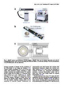
Abstract Objective This study aimed to investigate the optimal conditions for producing 68Ga-labeled tin colloid and the feasibility of 68 Ga-tin colloid positron emission tomography (PET) for visualization and evaluation of the phagocytic function of Kupffer cells (KCs) in vivo. Methods 68Ga-tin colloid was prepared by adding tin solution (1 mM, 0.2 mL) to 68Ga solution (1.0 mL), followed by pH adjustment with sodium acetate (1 M, 0.2 mL). Various labeling times were tested to find the optimal one. Colloid size was measured by filtering the solution through three-ply membrane filters (with pore sizes of 200, 3000, and 5000 nm), and radioactivity was measured in the whole filtrate and the filters using a gamma counter. The in vitro stability of the colloid was evaluated by the size measurement after incubation under ambient conditions for up to 60 min. PET scanning was performed for 30 min after intravenous administration of 68Ga-tin colloid solution (4 MBq) to healthy rats. Time-activity-curves for the liver, spleen, and blood pool were generated. Finally, liver uptake was compared before and after the establishment of KC-depletion and non-alcoholic steatohepatitis (NASH) rat models. Results Colloid size increased with increasing labeling time. After pH adjustment, the colloid sizes remained nearly unchanged. The optimal labeling time was determined as 30 min. PET imaging of healthy rats revealed that liver uptake of the 68Ga-tin colloid increased with increasing colloid size. In KC-depleted rats, liver uptake significantly decreased (n = 4, p
Data Loading...