Down-regulation of programmed cell death 5 by insulin-like growth factor 1 in osteoarthritis chondrocytes
- PDF / 356,700 Bytes
- 7 Pages / 595.276 x 790.866 pts Page_size
- 55 Downloads / 319 Views
ORIGINAL PAPER
Down-regulation of programmed cell death 5 by insulin-like growth factor 1 in osteoarthritis chondrocytes Chengqing Yi & Chunhui Ma & Zongping Xie & Guoqiao Zhang & Wangsheng Song & Xiaokai Zhou & Yun Cao
Received: 11 June 2012 / Accepted: 29 November 2012 / Published online: 16 January 2013 # Springer-Verlag Berlin Heidelberg 2013
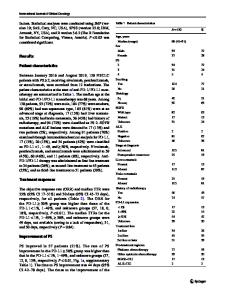
Abstract Purpose The aim of this study was to investigate the expression of insulin-like growth factor (IGF)-1 and programmed cell death 5 (PDCD5) in osteoarthritis chondrocytes, and to explore the potential correlation between them in the apoptosis process of osteoarthritis chondrocytes. Methods Patients with knee osteoarthritis were placed into four categories according to radiological staging. The mRNA and protein levels of IGF-1 and PDCD5 in osteoarthritis chondrocytes were respectively detected by quantitative reverse transcriptase polymerase chain reaction (qPCR) and western blotting. In addition, IGF-1 and PDCD5 protein expression in chondrocytes were also measured by immunohistochemistry. Apoptotic cells were measured by TUNEL staining. Results Both the mRNA and protein levels of IGF-1 were down-regulated, while the levels of PDCD5 were upregulated, and the mRNA and protein levels of IGF-1 were negatively correlated with those of PDCD5, respectively. The apoptotic cell was significantly increased in osteoarthritis chondrocytes compared with control. Importantly, the apoptosis rate was positively correlated with PDCD5 protein expression and negatively correlated with IGF-1 protein expression Conclusions We concluded that IGF-1 may down-regulate the expression of PDCD5 and thus inhibit the apoptosis of osteoarthritis chondrocytes. C. Yi (*) : C. Ma : G. Zhang : W. Song : X. Zhou : Y. Cao Department of Orthopaedics, Shanghai First People’s Hospital, No. 650 New Songjiang Road, Shanghai 201620, China e-mail: [email protected] Z. Xie Department of Orthopaedics, Shanghai Sixth People’s Hospital, Shanghai 200233, China
Introduction Osteoarthritis (OA) is a chronic arthropathy characterized by degenerative lesions of articular cartilage and peri-articular bone proliferation. OA is the most common arthritis and is also one of the major causes of joint pain in elderly people [1]. Several factors have been implicated in the pathogenesis of OA. For example, epidemiological studies show that OA may be caused by the joint effects of genetic and environmental factors [2, 3]. Loeser suggests articular chondrocytes exhibit an age-related decline in proliferative and synthetic capacity while maintaining the ability to produce proinflammatory mediators and matrix degrading enzymes, which contribute to the propensity to develop OA [4]. Recent studies have shown the presence of apoptotic as well as anti-apoptotic mechanisms in OA cartilage [5]. The OA cartilage has a higher proportion of apoptotic chondrocytes than normal tissue [6, 7]. Increased chondrocyte apoptosis may be related to up-regulated expression of both Akt and PKCα in human OA cartilage [8]. ADAM15, a disinte
Data Loading...