Electron Microscopy Study of Fatigue Crack Initiation in Ni 3 Al Single Crystals
- PDF / 2,554,243 Bytes
- 7 Pages / 420.48 x 639 pts Page_size
- 65 Downloads / 4,790 Views
ELECTRON MICROSCOPY STUDY OF FATIGUE CRACK INITIATION IN Ni3 Al SINGLE CRYSTALS L. M. Hsiung and N. S. Stoloff Materials Engineering Department Rensselaer Polytechnic Institute Troy, New York 12180-3590 ABSTRACT Transmission electron microscopy of single-slip oriented Ni3Al single crystals cyclically deformed at room temperature revealed a high density of dislocation dipoles and point defect clusters. The majority of the defect clusters were identified by weak-beam TEM to be of vacancy type. The observations of circular perfect dislocation loops, Frank loops, and spherical voids revealed the evidence of vacancy condensation during fatigue cycling at room temperature. Together with SEM observations of surface topography, it is suggested that fatigue crack initiation in Ni3 Al single crystals can be rationalized by formation of microvoids at PSB/matrix interfaces. INTRODUCTION The purpose of this research was to investigate the development of fatigue damage in Ni3 Al crystals. Particular emphasis has been placed upon the development of slip band surface topography, dislocation structures and role of point defect generation in crack initiation. EXPERIMENTAL PROCEDURE Single crystals with nominal chemical composition of Ni-24at%Al-O.25% were obtained from Pratt and Whitney Aircraft0 Div., United Technology Corp. The crystals were homogenized at 1150 C for 178 hrs in vacuum. Test specimens were prepared in the form of plates with gage dimensions of 3x3x9 mm prepared by electro-discharged0 machining (EDM). All specimens were annealed in vacuum at 1150 C for 50 hrs and electro-polished prior to testing. The orientation of stress axis is parallel to [2 13 17] as shown in Figure 1. This orientation is favorable for the primary octahedral slip system, (111)[701]. Strain-controlled tension-compression cyclic deformation tests, all beginning in tension, were performed on a closed loop machine. Single crystals were held by Wood's metal grips to facilitate alignment. Several tests were interrupted periodically to allow observation of surface slip traces by a Nomarski-type optical microscope or by scanning electron microscopy. The slip traces were determined by two surface trace analysis. Thin foils for transmission electron microscopy were prepared from gage sections of fatigued specimens. Dislocation structures were observed in a JEM-100CXII transmission electron microscope (operated at 100KV). The weak-beam imaging method was applied to study the dissociation of superdislocations and formation of point defects. Weak-beam images were recorded in the reflection vector g=+202 with sg_ 2.3xi0- nm1 corresponding to the g(3g) diffraction condition. Fracture surfaces were examined by SEM.
Mat. Res. Soc. Symp. Proc. Vol. 133. @1989 Materials Research Society
262
0
.5
=1
21-
0lii;.
-.- I1
_;0 103
10.110
Niimhr of cyr-Ies
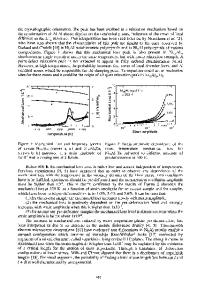
Figure 1 Cyclic-hardening curves for the [) 13 71] oriented Ni3 Al+B single crystals cycled at several strain amplitudes. EXPERIMENTAL RESULTS Cyclic Stress-Strain Response Cyclic strain-hardening curves f
Data Loading...