Immunoisolation of Nanoparticles Containing Endocytic Vesicles for Drug Quantitation
Cell-mediated nanoparticle delivery has recently emerged as an efficacious method of delivering therapeutic agents across physiological barriers. Use of cells as nanodelivery vehicles requires accurate assessment of their loading capacity and identificati
- PDF / 244,238 Bytes
- 6 Pages / 504.57 x 720 pts Page_size
- 10 Downloads / 288 Views
1
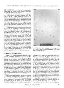
Introduction For over 20 years, nanoparticles (NP) have been researched for their use in drug delivery (1, 2). Drug-loaded NP have the potential to increase efficacy, reduce toxicity, and improve clinical outcomes of diseases. These nanoparticles tend to be designed to deliver drugs or other therapeutic compounds, such as protein or DNA, to specific cell populations. One of the important questions when researching drug-carrying nanoparticles is determining precisely how much drug the target cells are able to take up. Generally, this question is not difficult to answer. However, of even greater importance than how much drug the target cells are able to pick up is where within the cells are the nanoparticles being trafficked and stored.
Ari Nowacek and Irena Kadiu have contributed equally. Volkmar Weissig et al. (eds.), Cellular and Subcellular Nanotechnology: Methods and Protocols, Methods in Molecular Biology, vol. 991, DOI 10.1007/978-1-62703-336-7_5, © Springer Science+Business Media New York 2013
41
42
2
Ari Nowacek et al.
Materials Prepare all solutions using ultrapure water (prepared by purifying deionized water to attain a sensitivity of 18 MW cm at 25°C) and analytical grade reagents. Prepare and store all reagents at room temperature (unless indicated otherwise). Diligently follow all waste disposal regulations when disposing waste materials.
2.1 Conjugation of Magnetic Beads to Antibodies
1. PureProteome Protein A and Protein G Paramagnetic Beads (Millipore). 2. Antibodies to endosome surface markers of interest (see Note 1). 3. Bovine serum albumin fraction V (10%). 4. Sterile 1× phosphate-buffered saline (PBS). 5. Microcentrifuge tubes (1.7 mL). 6. Microcentrifuge tube tumbler rotator. 7. Magnetic separator rack. 8. Refrigerated tabletop centrifuge.
2.2 Cellular Treatment Components
1. Cells in culture (see Note 2). 2. Cell incubator. 3. Serum-free DMEM (or other appropriate serum-free culture medium). 4. Nanoparticles (see Note 3). 5. Sterile PBS.
2.3 Homogenization of NanoparticleLoaded Cells
1. Homogenization buffer: 10 mM HEPES–KOH, pH 7.2, 250 mM sucrose, 1 mM EDTA, and 1 mM Mg(OAc)2. 2. Cell scrapers. 3. Dounce homogenizer (7 mL). 4. 15 mL centrifuge tubes. 5. Refrigerated centrifuge.
2.4 Isolation of NanoparticleContaining Endosomes
1. Homogenate from Subheading 2.3). 2. Magnetic beads Subheading 2.1).
nanoparticle-treated with
attached
3. Magnetic separator rack. 4. Sterile PBS. 5. Refrigerated tabletop centrifuge.
cells
(from
antibodies
(from
Immunoisolation of Nanoparticles Containing Endocytic Vesicles…
2.5 Quantification of Drug Content by HPLC
43
1. HPLC-grade methanol. 2. Sonicator disruptor with probe tip. 3. Refrigerated tabletop centrifuge. 4. 0.5 mL microcentrifuge tubes. 5. HPLC autoinjector vials with low-volume inserts.
3
Methods
3.1 Conjugate Antibodies to Magnetic Beads
1. In a 1.7-mL microcentrifuge tube, combine 1 mL of 10% bovine serum albumin in PBS with 20 mL of magnetic bead slurry and 20 mg of antibody of interest (see Note 1). 2. Place tube
Data Loading...