Myocardial ASL perfusion reserve test detects ischemic segments in initial cohort of 10 patients with angiographic CAD
- PDF / 455,509 Bytes
- 2 Pages / 595.276 x 793.701 pts Page_size
- 54 Downloads / 337 Views
POSTER PRESENTATION
Open Access
Myocardial ASL perfusion reserve test detects ischemic segments in initial cohort of 10 patients with angiographic CAD Zungho Zun1*, Terrence Jao1, Ning Smith2, Padmini Varadarajan3, Ramdas G Pai3, Eric C Wong4, Krishna S Nayak1 From 2011 SCMR/Euro CMR Joint Scientific Sessions Nice, France. 3-6 February 2011 Objective This study sought to demonstrate the potential for myocardial arterial spin labeling (ASL) to identify the ischemic myocardial segments due to stenosis in coronary arteries as detected by X-ray angiography. Background Myocardial ASL is a technique for the assessment of myocardial blood flow (MBF) without contrast agents. It can be safely applied to patients with end-stage renal disease who are not candidates for first-pass imaging with contrast agents. Myocardial ASL perfusion imaging performed at rest and during adenosine stress provides perfusion reserve (MBFstress/MBFrest), which is a common indicator for the severity of coronary artery disease. In healthy myocardium, perfusion reserve is known to be approximately four [1].
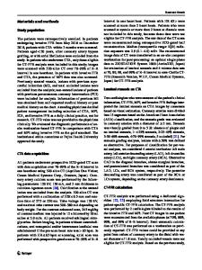
Results Ten of the twenty-nine patients were found to have significant stenosis on X-ray angiography. Table 1 contains the most ischemic myocardial segments in these ten patients as identified by two cardiologists using either X-ray angiogram or ASL perfusion reserve map independently. Based on McNemar’s test with Bonferroni correction, there was no significant difference between X-ray and ASL MRI in identifying ischemia in all six myocardial segments (p = 1.0000, 0.6170, 0.4795, 0.1336, 0.4795, and 0.4795). Figure 1 contains perfusion reserve maps acquired using myocardial ASL in these patients. The average standard deviation of physiological noise
Table 1 Most ischemic myocardial segments identified by X-ray angiograms and by ASL perfusion reserve maps Pts #
Methods Twenty nine patients were recruited from those scheduled for routine cardiac MR (CMR) and X-ray angiography. Myocardial ASL measurements were obtained from a single mid short-axis slice at rest and during adenosine infusion (dosage: 0.14 mg/kg/min) on a GE Signa 3T scanner. The ASL sequence was composed of flow-sensitive alternating inversion recovery (FAIR) tagging and balanced steady-state free precession (SSFP) imaging [2]. Perfusion reserve maps were generated in a standard short-axis view illustration by convolution with a Gaussian filter and resampling onto a polar coordinate [3].
1 University of Southern California, Los Angeles, CA, USA Full list of author information is available at the end of the article
X-ray angiography Worst lesion on angiogram
Ischemic myocardial segments
ASL MRI Ischemic myocardial segments
1
Proximal LAD 100%
Anterior
2
RCA 100%
Inferior, inferolateral Inferolateral
Anterior
3
LAD 90%
Anterior
4
LCS 90% (PDA)
Inferoseptal, Inferoseptal, inferior inferior, inferolateral
5
RCA (100%)
Inferoseptal, inferior Anteroseptal, inferoseptal, inferior
6
RCA (100%)
Inferoseptal, inferior Inferior
7
Distal RCA 80%
Inferior
Anterolat
Data Loading...