Nano- and Micro-Structural Evolution of UHMWPE by Ion Beam
- PDF / 390,261 Bytes
- 6 Pages / 612 x 792 pts (letter) Page_size
- 89 Downloads / 282 Views
1020-GG07-08
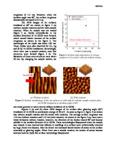
Nano- and Micro-Structural Evolution of UHMWPE by Ion Beam F. Calzzani1, B. Chhay1, R. Zimmerman1, A. Oztarhan2, and D. Ila1 1 Center for Irradiation of Materials, Alabama A&M University, 3900 Meridian Street, Normal, AL, 35762 2 Bioengineering, Ege University, Izmir Bornova, 35100, Turkey ABSTRACT It is important to produce uniform nano-patterns with no possibility of surface exfoliation on polyethylene devices used in medical and in aerospace industry. We studied the change in the surface morphology of polyethylene at nanoscale using MeV ion beam. We have investigated the change in the surface morphology before and after ion bombardment. We have made an attempt to change the morphology to produce a uniform surface with reduced cracks and reduced granularity. For this process we have chosen ultra-high-molecular-weight polyethylene (UHMWPE). Coupons of these materials were exposed to various fluences of MeV Ag+ ions. The surface morphology and the change in the chemical structure were studied using scanning micro Raman, FTIR and AFM. 1. INTRODUCTION Joint replacements are a very common surgical procedure used to restore function to cases that joints have been afflicted by major trauma or degenerating disease. Usually, those surgical approaches for joint replacement use a metal surface articulating against a component of UHMWPE. The use of UHMWPE became popular in total joint replacements due to its properties including biocompatibility, wear, friction, ductility and impact load resistance. However, wear particle-induced aseptic loosening of joint replacement prostheses remains a major cause of revision surgeries for the commonly used metal/ultra-high molecular weight polyethylene (UHMWPE) [1]. In addition to surface wear problems and despite the efforts of countless researchers to combat it, device-associated infection remains a major problem in medical care. Infection at medical replacements and indwelling catheters, for example, can result from contaminated disinfectants, from the hands of medical personnel, or as a result of self infection from a patient's own microflora. Such infections are not easily treated, since proliferating bacteria on the surface of the medical material can secrete a polysaccharide biofilm or "slime" difficult for systemic antibiotics to penetrate. Irradiation with Gamma rays [2] have been used extensively as the standard method of disinfection, however itís well known that this promotes undesirable oxidation under the prosthesis surface raising the wear production and causing a artificial aging. One alternative way of addressing device- related infection is to incorporate antimicrobial agents directly onto the surface of the device. Silver compounds (silver chloride or silver oxide) are a popular choice for infection-resistant coatings, but many commercially available silvercoated materials are of marginal effectiveness because the hydrophobic polymer matrix limits the silver ion concentration near the device surface.
In order to induce surface modifications in wear prope
Data Loading...