New Techniques Based on Ultrashort Laser Microsurgery for Use in Assisted Reproductive Technologies
- PDF / 730,469 Bytes
- 5 Pages / 612 x 792 pts (letter) Page_size
- 12 Downloads / 319 Views
Techniques Based on Ultrashort Laser Microsurgery for Use in Assisted Reproductive Technologies I. V. Ilinaa, *, Yu. V. Khramovab, M. A. Filatovb, A. D. Ivanovab, and D. S. Sitnikova aJoint
Institute for High Temperatures, Russian Academy of Sciences, Moscow, 125412 Russia bDepartment of Biology, Moscow State University, Moscow, 119991 Russia *e-mail: [email protected] Received June 18, 2020; revised July 10, 2020; accepted July 27, 2020
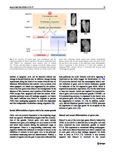
Abstract—Two noncontact techniques based on the use of ultrashort laser pulses are presented for assisted reproductive technologies. The first creates a unique alphanumeric code on an embryo’s envelope to simplify its identification during the period of preimplantation development. The second ensures the embryo will start to hatch at a prescribed site via laser-assisted drilling of its shell at the early blastocyst stage, where the inner cell mass and trophectoderm cells can be distinguished clearly. DOI: 10.3103/S106287382011012X
INTRODUCTION Sources of laser radiation have long been indispensable diagnostic and therapeutic tools in various fields of medicine. Assisted reproductive technologies (ARTs) are no exception. They are a relatively young but rapidly developing area of medicine, the main goal of which is to solve the problem of infertility in married couples. In ARTs, lasers are used to treat both male and female reproductive cells [1]. In the first case, laser sources are used to increase the motility of spermatozoa [2, 3] or immobilize them before an intracytoplasmic sperm injection [4, 5]. In the second, lasers are used mainly for microsurgery of the outer shell of an egg/embryo [6, 7], or individual microsurgical manipulations with embryonic cells (e.g., during a biopsy of the embryonic trophectoderm) [8, 9]. In this work, we consider new technologies for increasing the effectiveness and safety of existing ART techniques by using sources of ultrashort laser pulses for microsurgery of the zona pellucida of oocyte/preimplantation embryo. The first of these technologies relies on the action of ultrashort pulses (USP) on the zona pellucida of an egg/embryo to create micro-cuts on it in the form of an alphanumeric code, making it easy to identify each embryo during its preimplantation development and eliminate the risk of choosing the wrong embryo when transferring it to the uterine cavity. The second also includes laser microsurgery of the zona pellucida, but at later stages of preimplantation development (the blastocyst stage) when cell differentiation occurs and the inner cell mass (ICM) and trophoblast form. The clearly defined localization of the ICM at this stage enables us to form a hole in the membrane precisely in the best place and thereby con-
trol the beginning and place of embryo hatching. Hatching by “trophoblast forward” is thus preferable when a small number of trophectoderm cells must be selected for preimplantation genetic diagnosis (i.e., to perform a biopsy). The technology under development can therefore be referred to as that of controlled laser ha
Data Loading...