Nonlinear viscoelastic constitutive model for bovine liver tissue
- PDF / 6,925,528 Bytes
- 22 Pages / 595.276 x 790.866 pts Page_size
- 90 Downloads / 333 Views
ORIGINAL PAPER
Nonlinear viscoelastic constitutive model for bovine liver tissue Adela Capilnasiu1 · Lynne Bilston4,5 · Ralph Sinkus1,2 · David Nordsletten1,3 Received: 1 July 2019 / Accepted: 21 January 2020 © The Author(s) 2020
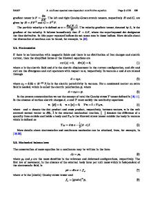
Abstract Soft tissue mechanical characterisation is important in many areas of medical research. Examples span from surgery training, device design and testing, sudden injury and disease diagnosis. The liver is of particular interest, as it is the most commonly injured organ in frontal and side motor vehicle crashes, and also assessed for inflammation and fibrosis in chronic liver diseases. Hence, an extensive rheological characterisation of liver tissue would contribute to advancements in these areas, which are dependent upon underlying biomechanical models. The aim of this paper is to define a liver constitutive equation that is able to characterise the nonlinear viscoelastic behaviour of liver tissue under a range of deformations and frequencies. The tissue response to large amplitude oscillatory shear (1–50%) under varying preloads (1–20%) and frequencies (0.5–2 Hz) is modelled using viscoelastic-adapted forms of the Mooney–Rivlin, Ogden and exponential models. These models are fit to the data using classical or modified objective norms. The results show that all three models are suitable for capturing the initial nonlinear regime, with the latter two being capable of capturing, simultaneously, the whole deformation range tested. The work presented here provides a comprehensive analysis across several material models and norms, leading to an identifiable constitutive equation that describes the nonlinear viscoelastic behaviour of the liver. Keywords Liver rheology · Biomechanics · Nonlinear mechanics · Viscoelasticity
1 Introduction Biomechanical characterisation of tissues is essential in medical research. New surgery techniques, implants or devices are being tested in silico, in vitro and in vivo (O’Toole et al. 1995; Marescaux et al. 1998; Rosen et al. 2008; Clin et al. 2010; Gonzalez-Blohm et al. 2015). In these tests, it is critical to know the liver’s response to a range of factors, such as puncturing, cutting, deformations and displacements. For diagnosis purposes, elastography is a technique that depends on the underlying tissue properties * Adela Capilnasiu [email protected] 1
Division of Biomedical Engineering and Imaging Sciences, King’s College London, London, UK
2
Inserm U1148, LVTS, University Paris Diderot, University Paris 13, 75018 Paris, France
3
Department of Biomedical Engineering and Cardiac Surgery, University of Michigan, Ann Arbor, USA
4
Prince of Wales Clinical School, University of New South Wales, Sydney, Australia
5
Neuroscience Research Australia, Sydney, Australia
in order to assess the presence of disease (Fovargue et al. 2018). In vivo magnetic resonance elastography (MRE) has shown that the existence of liver inflammation and fibrosis gives higher stiffness measurements (Huwart et al. 2006; Sinkus et al. 2018). However,
Data Loading...