Novel Murine Models of Cerebral Cavernous Malformations
- PDF / 5,488,801 Bytes
- 16 Pages / 595.276 x 790.866 pts Page_size
- 48 Downloads / 326 Views
ORIGINAL PAPER
Novel Murine Models of Cerebral Cavernous Malformations Matthew R. Detter1 · Robert Shenkar2 · Christian R. Benavides1 · Catherine A. Neilson1 · Thomas Moore2 · Rhonda Lightle2 · Nicholas Hobson2 · Le Shen2 · Ying Cao2 · Romuald Girard2 · Dongdong Zhang2 · Erin Griffin1 · Carol J. Gallione1 · Issam A. Awad2 · Douglas A. Marchuk1,3 Received: 10 March 2020 / Accepted: 6 July 2020 © Springer Nature B.V. 2020
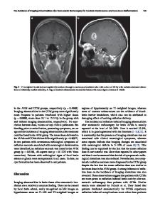
Abstract Cerebral cavernous malformations (CCMs) are ectatic capillary-venous malformations that develop in approximately 0.5% of the population. Patients with CCMs may develop headaches, focal neurologic deficits, seizures, and hemorrhages. While symptomatic CCMs, depending upon the anatomic location, can be surgically removed, there is currently no pharmaceutical therapy to treat CCMs. Several mouse models have been developed to better understand CCM pathogenesis and test therapeutics. The most common mouse models induce a large CCM burden that is anatomically restricted to the cerebellum and contributes to lethality in the early days of life. These inducible models thus have a relatively short period for drug administration. We developed an inducible CCM3 mouse model that develops CCMs after weaning and provides a longer period for potential therapeutic intervention. Using this new model, three recently proposed CCM therapies, fasudil, tempol, vitamin D3, and a combination of the three drugs, failed to substantially reduce CCM formation when treatment was administered for 5 weeks, from postnatal day 21 (P21) to P56. We next restricted Ccm3 deletion to the brain vasculature and provided greater time (121 days) for CCMs to develop chronic hemorrhage, recapitulating the human lesions. We also developed the first model of acute CCM hemorrhage by injecting mice harboring CCMs with lipopolysaccharide. These efficient models will enable future drug studies to more precisely target clinically relevant features of CCM disease: CCM formation, chronic hemorrhage, and acute hemorrhage. Keywords Cerebral cavernous malformation · CCM · Cavernous angioma · Stroke · Cerebral hemorrhage · Fasudil · Tempol · Vitamin D · Lipopolysaccharide
Introduction
Electronic supplementary material The online version of this article (https://doi.org/10.1007/s10456-020-09736-8) contains supplementary material, which is available to authorized users. Issam A. Awad and Douglas A. Marchuk contributed equally to this study. * Douglas A. Marchuk [email protected] 1
Department of Molecular Genetics and Microbiology, Duke University School of Medicine, Durham, NC 27705, USA
2
Neurovascular Surgery Program, Department of Neurosurgery, University of Chicago Medicine and Biological Sciences, Chicago, IL 60637, USA
3
James B Duke Professor, Department of Molecular Genetics and Microbiology, Duke University School of Medicine, Box 3175, Durham, NC 27710, USA
Cerebral cavernous malformations (CCMs), also known as cavernous angiomas, are clusters of dilated and brittle capillary-venous vessels that devel
Data Loading...