OCT angiography analysis of retinal vessel density in primary open-angle glaucoma with and without Tafluprost therapy
- PDF / 1,197,994 Bytes
- 8 Pages / 595.276 x 790.866 pts Page_size
- 87 Downloads / 278 Views
RESEARCH ARTICLE
Open Access
OCT angiography analysis of retinal vessel density in primary open-angle glaucoma with and without Tafluprost therapy Hannah Weindler*, Martin S. Spitzer, Maximilian Schultheiß and Robert Kromer
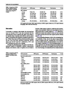
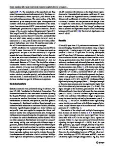
Abstract Background: Primary open-angle glaucoma (POAG) is a progressive neurodegenerative disease which leads to irreversible blindness. An elevated intraocular pressure (IOP) is considered to be the main risk factor for the disease progression. It is known that retinal blood flow is altered in POAG eyes. Tafluprost, a prostaglandin analogue which lowers the IOP, has shown to also improve the retinal blood flow in animals. Methods: The current study therefore evaluated the retinal vessel density in the peripapillary and macular region of POAG patients with normal IOP treated with topical Tafluprost (n = 20) compared to surgically treated patients with normal IOP (n = 22) using optical coherence tomography angiography (OCT-A). The retinal flow density was obtained after binarisation and evaluated in five sectors. Results: There was a significantly higher peripapillary flow density in all sectors in Tafluprost treated eyes when compared to post-surgery eyes. The flow density in the inferior sector of the superficial plexus in the macular region was also significantly higher in the Tafluprost group. Conclusions: These results indicate that Tafluprost not only lowers IOP, but may also enhance retinal blood flow in POAG patients with a normal IOP. Keywords: Primary open-angle glaucoma, Optical coherence tomography angiography, Tafluprost, Retinal flow density, Optic nerve head, Macular region
Background Primary open-angle glaucoma (POAG) is a leading cause for blindness worldwide [1, 2]. POAG shows no symptoms in early stages. Instead, patients present a slow, progressive deterioration of nerve fibre layer and increasing visual field losses. Glaucoma often affects both eyes. The pathophysiology of this chronic, multifactorial and neurodegenerative disease remains unclear [1]. An elevated IOP is the most crucial risk factor for POAG as well as disease progression [1]; hence, current treatment options focus on lowering it. However, * Correspondence: [email protected] Department of Ophthalmology, University Medical Center Hamburg-Eppendorf, Martinistraße 52, 20246 Hamburg, Germany
treating glaucomatous eyes is challenging and requires lifelong and thorough follow-up examinations. Within the therapeutic alternatives, surgical interventions aim to enable improved drainage of the aqueous humour through the implementation of a mechanical drain. Medicamentous treatment involves either reducing aqueous humour production or increasing drainage. However, the regulation of IOP in POAG does not ensure the stabilization of this irreversible disease and deterioration in the patient’s visual field remains possible. In addition to the mechanical pathophysiological theory of elevated IOP, retinal blood flow is reduced in glaucoma [3]. The damage caused by decreased retinal blood flow might contribute to the dev
Data Loading...