Painful Skin Nodules in Nephrotic Syndrome
- PDF / 454,962 Bytes
- 2 Pages / 595.276 x 790.866 pts Page_size
- 94 Downloads / 416 Views
PICTURE OF THE MONTH
Painful Skin Nodules in Nephrotic Syndrome Lesa Dawman 1
&
Karalanglin Tiewsoh 1 & K. Vinay 2 & Uma Nahar 3
Received: 11 March 2020 / Accepted: 8 May 2020 # Dr. K C Chaudhuri Foundation 2020
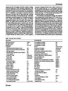
A 2 y 3 mo-old female child, with steroid dependent nephrotic syndrome was admitted with spontaneous bacterial peritonitis (SBP). The child was on daily prednisolone @ 2 mg/kg/d for 3 wk prior to admission as they did not come for follow up. For SBP, the child was started on stress dose steroids @ 0.5 mg/kg/d and IV antibiotics. After 10 d of admission, she developed tightening of skin with painful nodular skin lesions over bilateral upper and lower limbs and lower abdomen. On examination, multiple painful nodules over the thigh, legs, arms, forearm and abdomen were observed (Fig. 1a) and a possibility of panniculitis was kept. Skin biopsy showed intact epidermis with lymphocytic sprinkling of dermis; subcutaneous tissue showed septal infiltration by foamy macrophages with foci of calcification. Findings were consistent with histiocytic panniculitis (Fig. 1b-d). Prednisolone was reinitiated @ 2 mg/kg/d and tapered after remission. The tightening of skin improved within 2 wk; however skin nodules persisted. Few non-tender nodules were observed even at 20 mo followup. Currently, the child is on levamisole and tapering dose of prednisolone and her nephrotic syndrome is in remission. Steroids are the mainstay of therapy in children with nephrotic syndrome [1]. Post steroid panniculitis is a rare
* Lesa Dawman [email protected] 1
Department of Pediatrics, Post Graduate Institute of Medical Education and Research (PGIMER), Chandigarh 160012, India
2
Department of Dermatology, Venereology & Leprosy, Postgraduate Institute of Medical Education and Research (PGIMER), Chandigarh 160012, India
3
Department of Histopathology, Post Graduate Institute of Medical Education and Research (PGIMER), Chandigarh 160012, India
complication in children receiving high dose of systemic corticosteroids. It can occur within days to weeks of rapid tapering or stoppage of steroids. Majority are self-limiting and a small number of children require re-introduction of steroids. Clinical history and a skin biopsy, if necessary, helps in the diagnosis [2, 3]. The proposed mechanism is an increase in the normally elevated ratio of saturated to unsaturated fatty acids in the panniculus, leading to crystal formation due to rapid cessation of long-term corticosteroids [3]. Small, painful, occasionally pruritic nodules appear, mainly on the cheeks, arms and trunk, and areas prone to the greatest accumulation of fat during steroid treatment. Re-institution of corticosteroids has been shown to induce improvement of panniculitis; and selfresolving cases have also been observed [4]. Panniculitis, in general, can be encountered in children with vasculitic disorders, systemic infections, trauma or rarely with underlying pancreatic disease. In children with nephrotic syndrome, panniculitis have been encountered even without the use of st
Data Loading...