Photoluminescence of Eu-doped GaN
- PDF / 421,637 Bytes
- 9 Pages / 432 x 648 pts Page_size
- 5 Downloads / 437 Views
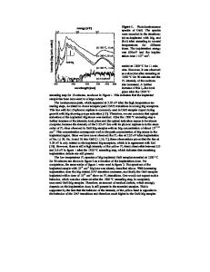
Photoluminescence of Eu-doped GaN K.P.O’Donnell, SUPA Dept. of Physics, University of Strathclyde, Glasgow G4 0NG, UK ABSTRACT This talk reviews work on the optical properties of Eu-doped GaN at the Semiconductor Spectroscopy laboratory of the University of Strathclyde. The principal experimental technique used has been lamp-based Photoluminescence/Excitation (PL/E) spectroscopy on samples produced mainly by high-energy ion implantation and annealing, either at low or high pressures of nitrogen, as described by Lorenz et al. [1]. These have been supplemented by samples doped in-situ either by Molecular Beam Epitaxy or Metallorganic Vapour Phase Epitaxy. Magnetooptic experiments on GaN:Eu were carried out in collaboration with the University of Bath. INTRODUCTION GaN doped with Europium, hereafter GaN:Eu, is currently of interest as a source of red light in prospective laser and light-emitting diodes. Despite quite a large recent effort, the defect structure, excitation mechanism and even the basic spectroscopy of GaN:Eu, in common with other rare-earth ion and III-nitride combinations, is not well understood. EXPERIMENTAL DETAILS Photoluminescence/Excitation (PL/E) spectroscopy at the University of Strathclyde employs a home-built apparatus (figure 1) which couples a 1 kW Xe arc lamp with a ¼ m JobinYvon monochromator as the excitation source and a McPherson 2/3 m spectrometer with a cooled photomultiplier tube (PMT) for analysis and detection of the sample fluorescence. Samples, mounted in an optical cryostat on an x-z stage, are cooled by a 2-stage closed-cycle Helium refrigerator to a base temperature of ~15 K. An Oxford Intruments temperature controller uses a strip heater to elevate the sample temperature in the range 15 – 300 K.
Figure 1: PL/E spectroscopy apparatus.
101
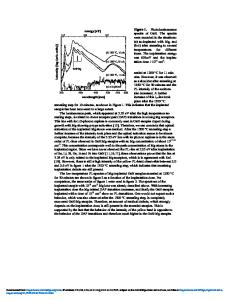
The PL/E system can be used with a fixed excitation wavelength to obtain photoluminescence (PL) spectra by recording the amplified PL signal as a function of the emission wavelength. Excitation spectra monitor the PL signal at a particular wavelength of emission while scanning the excitation spectrometer wavelength. A digital interface controls the monochromators by means of stepper motors and the spectra are recorded and displayed by computer. The dispersion of the emission monochromator (0.6 nm per mm of slit with a 2400 line mm-1 grating) allows good resolution of sharp emission lines, which are typically ~0.2-3 nm wide (FWHM) at ~620 nm (i.e. < 0.5 meV), while the excitation monochromator is usually set to provide an adequate sample illumination of order 1 mW cm-2 in a 10 meV bandpass. Preliminary room temperature cathodoluminescence (CL) spectroscopy complements the PL/E measurements. In general CL uses very much higher excitation densities than PL/E. EXPERIMENTAL RESULTS AND DISCUSSION MOCVD samples By far the brightest GaN-based red-light emitters examined to date have been MOCVDdoped samples produced at Osaka University by Fujiwara and co-workers [2]. We begin with an overview of the cathodoluminescence (CL) and PL/E of these sampl
Data Loading...