Photoquenching of Persistent Photoconductivity in N-Type GaN
- PDF / 431,301 Bytes
- 6 Pages / 414.72 x 648 pts Page_size
- 18 Downloads / 368 Views
EXPERIMENTAL The GaN samples used in this study were grown by metal organic chemnical vapor deposition (MOCVD). The filns with a thickkness between 3 and 3.4 jism were grown on sapphire substrates. The epilayers were unintentionally doped ni-type with carrier concentrations between 531 Mat. Res. Soc. Symp. Proc. Vol. 482 01998 Materials Research Society
5
a)
,4,0
b)
I light off
GaNT=77K X=325nm
3,9 C '
c 4-4
5E C8 2""
light off
E C
a)ca,
- 33-
3,8 "-Uo L -
=3 02
o o0
2,0
2,4
2,8
Energy [eV]
3,2
3,6
2
0
V
light on
0
1
Time [1 04s]
2
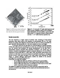
Figure 1: (a) Low temperature PL spectrunm of GaN filn with a room-temperature carrier concentration of Nj = 1.7 x 10¹⁸ cn- 3 . (b) Typical buildup and decay transients of the conductivity at 77K and 302K (excitation wavelength 388nm). 1 x 10¹⁷ and 1.7 x 10¹⁸ ci- 3 . Photoluminescence (PL) spectra of the films were recorded at 5K using excitation light of 325nm. Coplanar ohmic Ti/Al contacts in Hall geometry were formed on the GaN surface for conductivity measurements. For the photoexcitation studies we used monochromatic light from a Xe-lamp dispersed by a 0.25m-monochromator. The photon flux on the samples was of the order of 1011 - 1012 s'. For some experiments the samples were illuminated by light emitting diodes (LEDs) radiating at different wavelengths, with a full-width-half-maximum of -25ini simultaneously with the monochromatic illumination. To study the PPC effects the samples were kept in the dark at room temperature (--300K) before they were cooled to their respective measurement temperature (77-300K). The saturation photoconductivity levels under different illumination conditions were measured in a single cooling cycle at 100K. RESULTS AND DISCUSSION We checked the quality of the GaN films by measuring the low temperature photoluminescence spectra of the films. The spectra exhibit a strong and very narrow near band-edge peak with a half width of 2.8mieV, as presented in Fig. La. The yellow emission band, around 2.2eV, which is often prominent in films with inferior film quality, is very weak for our samples. Its intensity is only about 1.3x 10- of the intensity of the donor bound exciton peak. Comparable PL spectra have been found for several samples with different carrier densities and nobilities. In Fig. 11) we present typical PPC excitation and dlark decay transients for a sample at room temperature (RT) and at 77K. The PPC is induced by subbandgap illumination (A =388nm) as discussed in R ef.[7]. The PPC' is more pronounced at low temperatures T. The build-up time depends on the photon flux P and the optical ionization cross section aopt,
which was used as a measut'e for the optical ionization energy of the deep traps involved [7]. 532
1.2
1.0 (D 0.8 -CU
0 0.6.. 0
illumination
0.4 -
X=388nm
I,
0.0
I
0.5
,
I
1.0
dark •
I
1.5
W..
I
dark
2.0
,
H111
2.5
dark l
3.0
Time [103 s] Figure 2: Decay transient of the PPC at 200K when the sanlple was illunminated by 780nmn light during the indicated time intervals. After the
Data Loading...