Pre-eminence of the Indirect Channel in the Resonant Inverse Photoelectron Spectroscopy of Cerium Oxide
- PDF / 317,950 Bytes
- 6 Pages / 432 x 648 pts Page_size
- 90 Downloads / 287 Views
Pre-eminence of the Indirect Channel in the Resonant Inverse Photoelectron Spectroscopy of Cerium Oxide J. G. Tobin1,*, S.-W. Yu1, B.W. Chung1, and G.D. Waddill2 1
Lawrence Livermore National Laboratory, Livermore, CA, USA Missouri Univ. of Science and Technology, Dept of Physics, Rolla, MO 65401 *Corresponding Author, [email protected]
2
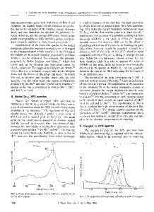
ABSTRACT A strong resonance in the inverse photoelectron spectroscopy (IPES) of cerium oxide was reported recently. Here, it is shown that dominance of the indirect channel of the resonant inverse photoelectron spectroscopy (RIPES) is so complete that the photon energy dependence can be explained in terms of emission associated with a single photon energy. INTRODUCTION Soft X-ray spectroscopy provides a powerful approach for the interrogation of the electronic structure of materials and surfaces. Three particularly important variants are inverse photoelectron spectroscopy (IPES) [1], X-ray emission spectroscopy (XES) [1], and X-ray absorption spectroscopy (XAS) [2]. Examples of the data for each are shown in Figure 1. Diagrams of the underlying processes are illustrated in Figure 2. In the case of inverse photoelectron spectroscopy, also known as Bremstrahlung Isochromat Spectroscopy (BIS) at high energies, it is possible to operate under resonant conditions (RIPES), where a second, indirect channel can contribute to the intensity, analogous with resonant photoelectron spectroscopy (RESPES) [3]. The indirect channel involves going through a core level with the concomitant requirement that the direct and indirect channels end with the same final state [1,3]. In the case of the Ce3d levels, of cerium oxide, the resonance is extraordinarily strong and the indirect channel is overwhelmingly pre-eminent, as will be shown below. EXPERIMENT The XES and RIPES experiments were carried out at Lawrence Livermore National Laboratory (LLNL), as described elsewhere [1]. The XAS measurements were made on Beam-line 8 at the Advance Light Source (ALS) at Lawrence Berkeley National Laboratory (LBNL), in Berkeley, CA [2]. As discussed in detail in Reference 1, the stoichiometry of the cerium oxide is unknown.
289
Figure 1. (Left) The XAS, XES and RIPES at hv = 881 eV are plotted here. (Top) The XAS of cerium metal, cerium oxide on cerium metal and reference spectra of a thin film of cerium oxide [2]. The instrumental broadening of the XAS is on the scale of 0.1 eV to 0.2 eV, allowing for the observation of substantial fine structure. TEY is total electron yield. TYF is total fluorescence yield. (Bottom) The XES and RIPES of Ce oxide/Ce metal sample [1]. The instrumental resolution is on the scale of about 2 eV for the RIPES and less than 2 eV for the XES. This broader instrumental band-pass contributes to the loss of fine structure relative to XAS. Figure 2. (Below) This is a schematic illustrating the essentials of the processes for XAS, XES and RIPES of the Ce3d5/2 and Ce3d3/2 levels. Non-resonant IPES or BIS would correspond to the direct channel in RIPES. HF is the energy of the hole to be fi
Data Loading...