Primary small cell type of non-Hodgkin lymphoma of the colon: a case report
- PDF / 1,417,322 Bytes
- 3 Pages / 595.276 x 790.866 pts Page_size
- 48 Downloads / 303 Views
(2020) 14:157
CASE REPORT
Open Access
Primary small cell type of non-Hodgkin lymphoma of the colon: a case report Eyal Meir1,2, Chovav Handler1,2, Uri Kaplan1,2, Doron Kopelman1,2 and Ossama A. Hatoum1,2*
Abstract Introduction: Primary lymphoma of the colon is exceedingly rare and comprises 0.2–1% of all colon tumors. The most common subtype of lymphoma in the colon is non-Hodgkin lymphoma. Symptoms are often nonspecific, and treatment varies between chemotherapy alone and a combination of surgery and chemotherapy. Case presentation: We describe a case of a Ashkenazi Jew patient who presented in the typical way that carcinoma of the colon might present but turned out to have a very rare type of tumor in both its histology and its location. Conclusion: There was apparent discordance between the relative bulkiness and gross appearance of the tumor with the unrevealing result of the biopsies, demanding a high level of suspicion as to the actual presence and possible type of such a tumor in the future. Keywords: Case report, Colon, Lymphoma
Introduction Primary lymphoma of the colon is exceedingly rare and comprises 0.2–1% of all colon tumors [1–4]. The most common subtype of lymphoma in the colon is nonHodgkin lymphoma (NHL) [5]. Though the most common site for secondary spread of lymphoma is the gastrointestinal (GI) tract, primary lymphoma of the GI tract accounts for only 10–15% of all lymphomas. The most common GI location for primary lymphoma is the stomach 25-50%, followed by the small intestine (20– 30%). The colon and rectum account for the remaining 10–20% [6, 7]. Symptoms are often nonspecific, and treatment varies between chemotherapy alone and a combination of surgery and chemotherapy [8]. Case presentation A 57-year-old Ashkenazi Jew woman, who aside from iron deficiency anemia was relatively well, with no family or personal history of malignancy, was admitted to our * Correspondence: [email protected] 1 Department of Surgery B, Emek Medical Center, Afula, Israel 2 Rapaport Faculty of Medicine, Technion-Israel Institute of Technology, Haifa, Israel
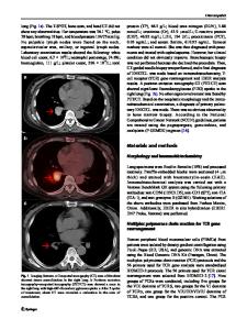
department of general surgery for treatment of her transverse colon tumor. Four months prior, she had begun experiencing periumbilical abdominal pain hematochezia, and she had a 10-kg weight loss. Upon physical examination, no masses were palpated, and there were no other pathologic findings. She underwent a colonoscopy, which revealed a large mass that involved nearly the whole circumference of the colon and seemed to be adjacent to the cecum. Biopsies were taken that failed to demonstrate any colonic pathology. She proceeded to undergo computed tomography (CT) of the chest and abdomen that demonstrated a huge mass that occupied the whole colonic lumen and caused a colocolic intussusception (Fig. 1). Considerable mesenteric lymphadenopathy was seen with nodes up to 28 × 21 mm in diameter and was deemed to be evidence of positive tumoral lymph node involvement (Fig. 2). No inguinal, pelvic, retroperitoneal, or other lymphadenopathy was seen. Considering t
Data Loading...