Pulmonary benign metastasizing leiomyoma: a case report and review of the literature
- PDF / 252,528 Bytes
- 4 Pages / 595.28 x 793.7 pts Page_size
- 115 Downloads / 285 Views
CASE REPORT
WORLD JOURNAL OF SURGICAL ONCOLOGY
Open Access
Pulmonary benign metastasizing leiomyoma: a case report and review of the literature Yili Fu, Hui Li*, Bo Tian and Bin Hu
Abstract Pulmonary benign metastasizing leiomyoma characterized by the growth of uterine leiomyoma in the lung is a very rare disease. We herein report the case of a 46-year-old asymptomatic woman who underwent a total abdominal hysterectomy for her multiple uterine leiomyomas 5 years ago, with the presence of multiple shadows in her chest roentgenogram during the regular check-up. Chest computerized tomography (CT) showed multiple solitary nodules in both lungs. Video-assisted thoracoscopic surgery with a wedge resection of the lesion was performed. Histopathologically, the pulmonary nodule was composed of benign smooth muscle cells and demonstrated low mitotic activity and absence of necrosis. Immunohistochemical staining for smooth muscle actin (SMA) and Desmin were extremely positive. CD10, CD117 and S-100 were negative in the tumor cells. Positive immunoreactivity for estrogen receptor (ER) and progesterone receptor (PR) were detected. The pathological diagnosis was pulmonary benign metastasizing leiomyoma. Keywords: Lung, Leiomyoma, Benign lesion, Metastases
Background Pulmonary benign metastasizing leiomyoma is a very rare disease characterized by the growth of uterine leiomyoma tissue in the lung [1]. In most cases, there is a previous history of total abdominal hysterectomy for uterine leiomyoma. However, the pathogenesis of this disease has not yet been elucidated. Case presentation A 46-year-old Chinese woman, who was a nonsmoker, was admitted for further examination of scattered shadows in both lungs that were found incidentally on the chest roentgenogram during a regular checkup in 2011. A chest computerized tomography (CT) was subsequently performed. The patient’s past medical history showed a diagnosis of multiple uterine leiomyoma in 2005 for which a total abdominal hysterectomy had been performed. The pathology revealed a uterine smooth muscle tumor with vascular invasion. During the 5-year follow-up, no recurrence of the benign metastasizing leiomyoma was detected. The clinical examination was unremarkable, and the results from routine laboratory * Correspondence: [email protected] Department of Thoracic Surgery, Beijing Chao-Yang Hospital, Capital Medical University, Beijing 100020, People’s Republic of China
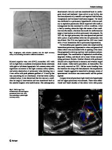
examinations were normal. CT demonstrated multiple solitary nodules in both lungs with maximum diameters of 2 cm (Figure 1A). Positron emission tomography (PET/CT) did not show abnormal fluorodeoxyglucose uptake with the suspicion of multiple nodules, and the SUV value was about 2 (Figure 1B). The diagnosis at the time of admission was suspected pulmonary metastasizing leiomyoma. In August 2011, the patient underwent right videoassisted thoracoscopic surgery with wedge resection of the lesion. One of the lesions from the right lower lobe was resected for pathological diagnosis. There were multiple small no
Data Loading...