Pulsatile Swelling of Umbilicus in a Cyanotic Neonate
- PDF / 323,964 Bytes
- 2 Pages / 612 x 792 pts (letter) Page_size
- 16 Downloads / 262 Views
Pulsatile Swelling of Umbilicus in a Cyanotic Neonate
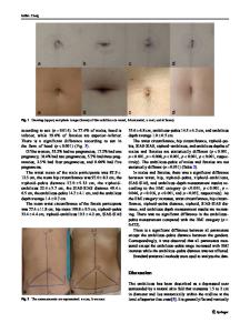
(Fig. 1c). Defect in anterior diaphragm was present, but there was no associated sternal defect. In view of atrial flutter, patient was started on propranolol and digoxin, and good heart rate control was achieved. After discussion with the cardiac surgical team, it was decided to close the left ventricular diverticulum, the cardiac lesion to be addressed later, since the oxygen saturation of the baby stayed above 85%. Left anterolateral thoracotomy was done and the fistulous tract arising from anterior most part of the left ventricular apex was identified. It was double clamped and divided and both ends were sutured. Defect in diaphragm was closed. The umbilical swelling was excised and the skin repaired. Patient had a smooth post-operative course and recovered well. The cardiac rhythm reverted to sinus rhythm on postoperative day 6. He was discharged on ninth post-operative day. Digoxin was stopped at six-week follow-up. Currently, at one-year follow up, baby is doing well, his oxygen saturation is 80% and he is in sinus rhythm. He is planned for Glenn surgery in view of non-committed muscular VSD, and is awaiting the same.
C
antrell syndrome is a rare, usually lethal, congenital malformation [1]. In the complete form, five anomalies exist, namely a midline supra-umbilical abdominal wall defect, a sternal defect, an anterior diaphragmatic defect, a diaphragmatic pericardial defect and a congenital heart defect. However, the extent of individual defects and their combination varies considerably; broad spectrum of associated cardiac abnormalities have been reported in most cases. We describe a neonate presenting with a pulsatile umbilical swelling and cyanosis since birth, later confirmed to be due to Cantrell syndrome. A full term male neonate with uneventful antenatal and perinatal course, born to a primigravida mother by normal vaginal delivery, was noted to have a pulsatile umbilical mass immediately after birth. Antenatal second trimester sonographic scans were reported normal, but detailed anomaly scan was not done. Baby was seen at our institute on day seven of life. He was feeding well, had a capillary filling time
Data Loading...