1014 Spiral imaging at 3 T for the measurement of endothelial-dependent coronary arterial function
- PDF / 665,148 Bytes
- 3 Pages / 610 x 792 pts Page_size
- 31 Downloads / 282 Views
BioMed Central
Open Access
Meeting abstract
1014 Spiral imaging at 3 T for the measurement of endothelial-dependent coronary arterial function Glenn Hirsch*, Sebastian Kelle, Allison Hays, Gary Gerstenblith, Michael Schar, Robert Weiss and Matthias Stuber Address: Johns Hopkins University, Baltimore, USA * Corresponding author
from 11th Annual SCMR Scientific Sessions Los Angeles, CA, USA. 1–3 February 2008 Published: 22 October 2008 Journal of Cardiovascular Magnetic Resonance 2008, 10(Suppl 1):A139
doi:10.1186/1532-429X-10-S1-A139
Abstracts of the 11th Annual SCMR Scientific Sessions - 2008
Meeting abstracts – A single PDF containing all abstracts in this Supplement is available here. http://www.biomedcentral.com/content/pdf/1532-429X-10-S1-info.pdfThis abstract is available from: http://jcmr-online.com/content/10/S1/A139 © 2008 Hirsch et al; licensee BioMed Central Ltd.
Background Invasive measures of endothelial-dependent coronary artery vasoreactivity predict cardiovascular event rates [1,2]. We previously demonstrated that endothelialdependent coronary vasoreactivity (EDCV) can be measured non-invasively by combining 3 T coronary MRI and isometric handgrip exercise [3]. Although coronary vasoreactivity is likely a stronger a predictor of cardiac events than similar measures in peripheral arteries such as the brachial, there are several coronary MRI techniques in use but no currently standardized protocol for non-invasively assessing EDCV. We assessed EDCV with 3 T MRI at regular time intervals before, during and after isometric handgrip exercise using two different cine pulse sequences, gradient echo (TFE) and spiral.
Methods Fifteen healthy, fasting subjects (8 female), age 28.9 ± 5.0 years were placed prone in a 3 T MRI scanner equipped with a 6-element cardiac coil. Thirteen subjects had adequate scans for analysis. After scout scan determination of proximal coronary orientation, a dedicated TFE cine sequence (FOV = 350 mm, slice thickness 8 mm, Matrix = 400, spatial resolution = 0.8 × 0.91 × 8 mm, temporal resolution = 47 ms, TR = 5.8 ms, TE = 3.5 ms, flip angle = 15°, BH duration = 15 sec, TFE factor = 8, SENSE, no fatsat, triggering = retrospective) perpendicular to the coronary artery of interest was acquired. A spiral sequence (FOV 300 mm, Matrix = 336, TE = 2.1 ms, Flip angle = 20°, BH duration = 15 sec, acquisition window = 17 ms,
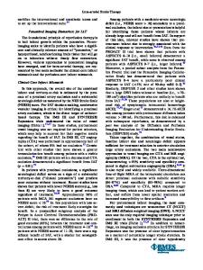
temporal resolution = 22.7 ms, spatial resolution 0.89 × 0.89 × 8.0 mm, SENSE = no, fatsat = spectral spatial, triggering = prospective) was then performed at the same anatomical level. Alternating TFE and spiral images were obtained at regular time intervals at baseline, during 4 minutes of continuous isometric handgrip exercise at 30% of each subject's maximum, and for up to 12 minutes of recovery. The images were analyzed for cross-sectional area changes using automated software employing fullwidth-half-maximum criteria (Cine version 3.15.17), see Figure 1. The total number of analyzable frames was counted by consensus reading for each method. Result
Data Loading...