Accuracy of various fluoroscopic landmarks for determination of midline implant placement within the cervical disc space
- PDF / 744,581 Bytes
- 6 Pages / 595.276 x 790.866 pts Page_size
- 41 Downloads / 259 Views
ORIGINAL ARTICLE
Accuracy of various fluoroscopic landmarks for determination of midline implant placement within the cervical disc space Peter B. Derman1 · Erik Waldorff2 · Nianli Zhang2 · Ram Haddas1 Received: 24 July 2020 / Revised: 24 July 2020 / Accepted: 10 October 2020 © Springer-Verlag GmbH Germany, part of Springer Nature 2020
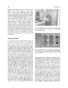
Abstract Purpose The traditional teaching has been that proper function of a cervical disc replacement is dependent upon appropriate placement, which includes centering the device in the coronal plane. The purpose of this study was to identify the most reliable anatomical landmark for determining midline placement of an implant within the cervical disc space under fluoroscopy. Methods Digital fluoroscopy images were taken for each cervical level at 0 °, 2.5 °, 5 °, 7.5 °, 10 °, and 15 ° from the midaxis by rotating the C-arm beam of six cadavers. Thin-slice CT scanning of the same levels was subsequently performed. Three independent reviewers measured the distance between anatomic structures: (a) tip of the right uncinate; (b) medial border of the right pedicle; and (c) center of the spinous processes for different x-ray angles across cervical levels C3–7. Results Both the uncinate and pedicle demonstrated superior overall accuracy to that of the spinous process (p ≤ 0.02) at all angles except at 0 ° for the pedicle where the difference was not statistically significant. Overall (pooled C3–7), the accuracy of the uncinate did not differ significantly from that of the pedicle at any fluoroscopic angle. The center of the spinous process measurement was particularly sensitive to deviations from the perfect anteroposterior fluoroscopy image. Conclusions The results of this investigation suggest that the tip of the uncinate and the medial border of the pedicle are more accurate measures of midline in the cervical spine than the center of the spinous process and are less susceptible to inadvertent off-axis imaging. Keywords Cervical disc replacement · Cervical spondylosis · Anterior cervical discectomy and fusion
Introduction Cervical disc replacement (CDR) is an increasingly popular motion-preserving alternative to fusion in appropriately selected patients with cervical spondylosis [1]. Compensatory motion and subsequent degeneration at adjacent levels may be minimized when segmental motion is maintained via arthroplasty. Studies have consistently demonstrated clinical and radiographic results that are equal or superior to those of anterior cervical discectomy and fusion (ACDF) [2]. The traditional teaching has been that proper function of a CDR device is dependent upon appropriate placement, which includes centering the implant in the coronal plane
* Ram Haddas [email protected] 1
Texas Back Institute, 6020 West Parker Road, Plano, TX 75093, USA
Orthofix Medical, Inc, Lewisville, TX, USA
2
[3]. In the lumbar arthroplasty literature, there is evidence to suggest that suboptimal implant placement is associated with inferior range of motion and clinical outcomes [4].
Data Loading...