Addition to: novel harvesting technique of no-touch saphenous vein graft using THUNDERBEAT
- PDF / 732,222 Bytes
- 2 Pages / 595.276 x 790.866 pts Page_size
- 84 Downloads / 243 Views
LETTER TO THE EDITOR
Addition to: novel harvesting technique of no‑touch saphenous vein graft using THUNDERBEAT Azumi Hamasaki1 Received: 5 January 2020 / Accepted: 1 March 2020 © The Japanese Association for Thoracic Surgery 2020
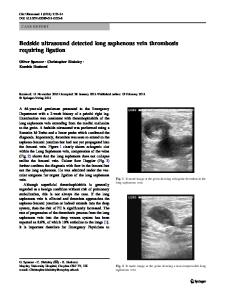
Keywords Coronary artery bypass grafting · No-touch saphenous vein graft · Harvesting technique · THUNDERBEAT After the “novel harvesting technique of no-touch saphenous vein graft using THUNDERBEAT” was published [1], the technique has been further improved. I would like to introduce improvements. The key point of improvement is to switch the harvesting method based on the anatomical features [2] of the saphenous fascia (SF) in front of the saphenous vein (SV), the muscular fascia (MF) in posterior, and the saphenous compartment (SC) sandwiched between the two fasciae. The SC contains only the SV, its branches, the saphenous nerve, and the surrounding adipose tissue. Short-axis image of the SC is likened to the eye of Horus (Fig. 1) in ancient Egypt. With this improvement, harvesting can be performed faster, and it is possible to harvest the no-touch SV with an appropriate amount of surrounding adipose tissue. This improvement prevents excessive defect of adipose tissue of the SC, thus making wound closure easier and reducing the risk of wound trouble. The improved sampling method is described below and shown in Fig. 2. Cut the skin and divide the subcutaneous fat tissue by an electrocautery down to the surface of the SF in front of the SC. After exposing the SF over the entire length of the SV, mark the SV with dye. Then, mark the planned incision lines on both sides approximately 5 mm away from the SV, like a railway track using an electrocautery. The SF and surrounding adipose tissue are coagulated and incised together just above the MF using the THUNDERBEAT Open Fine Jaw (Olympus Medical Systems Corp., Tokyo, Japan) along the planned incision lines. To make harvest easier, rotate the SC containing the SV like a roll after both
Fig. 1 A short-axis ultrasound image of the saphenous compartment (SC) resembling the eye of Horus is demonstrated. The saphenous vein (asterisk) is surrounded by adipose tissue (dagger). The front and back of the SC are covered by the saphenous fascia and muscular fascia, respectively. On the upper left corner is the eye of Horus symbol
sides of the SC are coagulated and incised. The back side of the SC is easily peeled off because there is nothing to pay attention to damage on the back side of the SV. I introduced the improvement points of the harvesting method. I hope this helps readers.
* Azumi Hamasaki hamasaki‑[email protected] 1
Second Department of Surgery, Yamagata University Faculty of Medicine, 2‑2‑2 Iida‑Nishi, Yamagata 990‑9585, Japan
13
Vol.:(0123456789)
General Thoracic and Cardiovascular Surgery
Fig. 2 Schematic illustrations of the harvesting method of the no-touch saphenous vein (SV) based on the anatomical features of the saphenous fascia (SF), the muscular fascia (MF), and the saphenous compartment (SC). a After exposing the
Data Loading...