Analyzing the Mesoscopic Structure of Pericellular Coats on Living Cells
- PDF / 8,057,243 Bytes
- 6 Pages / 612 x 792 pts (letter) Page_size
- 12 Downloads / 340 Views
1274-QQ02-03
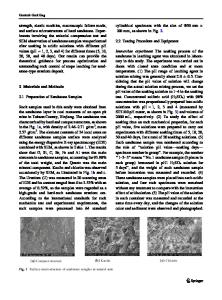
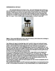
Analyzing the Mesoscopic Structure of Pericellular Coats on Living Cells H. Boehm1, T. A. Mundinger1, V. Hagel1, C. H. J. Boehm1, J. E. Curtis1,2, J. P. Spatz1 1 Max-Planck-Institute for Metals Research, Department New Materials & Biosystems, Heisenbergstr. 3, 70569 Stuttgart, Germany & University of Heidelberg, Department of Biophysical Chemistry, INF 253, 69120 Heidelberg, Germany. School of Physics, Georgia Institute of Technology, 837 State Street, Atlanta, GA, USA ABSTRACT We employed passive particle-tracking microrheology to map the micromechanical structure of the hyaluronan-rich pericellular coat enveloping chondrocytes. Therefor we exploited the technique’s position sensitivity to gain radial information on the coat. We observed a linear increase in viscoelasticity from the coat’s rim towards the cell membrane. This gradient corresponds to hyaluronan concentration profiles observed in confocal fluorescent microscopy with small, specific hyaluronan markers. These results suggest that the structural basis of the pericellular coat is formed by grafted hyaluronan of different effective lengths stretched out by a homogenous decoration with hyaladherins such as aggrecan. The different effective lengths could be caused either by different lengths of the HA chains or by “side-on” attachments within the chain. Remarkably, the hyaluronan-rich coat increases the viscosity of the pericellular space only by about a factor of about two at 100 and at 20 Hz compared to pure media and an increasing elastic component is observed. Both the viscoelasticity as well as the hyaluronan concentration decrease linearly or slightly exponential from the cell membrane towards the PCC’s rim. These observations could be obtained on living cells exploiting this unintrusive measurement techniques. INTRODUCTION Hyaluronan, a linear muco-polysaccharide forms the vital backbone of the highly hydrated pericellular coats (PCC) enveloping a variety of mammalian cells.1 The hyaluronan (HA) chains of several micrometer lengths2,3,4 serve as polyvalent templates binding several different proteins, called hyaladherins.5,2 Some of these, such as CD44 or RHAMM, are membrane proteins grafting the hyaluronan to the cell surface.3 Others like aggrecan are required for the formation of the PCC6 and might serve to stiffen the hyaluronan even at very low concentrations.7 The interplay of HA’s polymer properties and its structural organization and interaction with hyaladherins influence its micromechanical features and thus the adhesion process,8 proliferation, motility and embryogenesis9. First averaged elasticity values could be obtained with strain apparati10,11 and micropipet aspiration12 and could even distinguish between arthritic and normal human chondrocytes13. Local measurements at different areas of the cell can be performed with atomic force microscopy.14,15,16 However, so far all of the applied rheometric techniques can not gather information about the micromechanical structure of the PCC, which determines the PCC’s interaction wit
Data Loading...