Anatomy and Physiology of the Vitreo-macular Interface
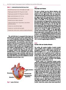
The vitreous occupies four fifths of the volume of the eyeball and consists of collagen fibers and hyaluronic acid. These components maintain a clear matrix with viscoelastic properties resulting in outward mechanical forces responsible for retinal attach
- PDF / 363,492 Bytes
- 7 Pages / 504.57 x 720 pts Page_size
- 6 Downloads / 285 Views
Anatomy and Physiology of the Vitreo-macular Interface Amitha Domalpally, Sapna Gangaputra, and Ronald P. Danis
3.1
History
The vitreous humor (vitreus, glassy; humor, fluid) has been recognized as a distinct anatomic structure since the ancient times (Duke-Elder and Wybar 1961). The first attempts to systematically describe the vitreous anatomy were seen in mideighteenth-century observations (Duke-Elder and Wybar 1961). Microscopic studies on the vitreous began in mid-nineteenth century leading to differing interpretations about the structure of the vitreous. Much of the controversy was related to the difficulties with microscopic preparation of the vitreous due to its high water content and difficulties with staining and fixation, which led to highly variable specimen appearances (Wolf 1968). The fibrillar structure of the vitreous seen on slit lamp biomicroscopy further added to speculation on vitreous structure. Theories by notable anatomists were advanced, such as the alveolar theory by Demours, the lamellar theory by Zinn, the radial theory by Hannover, and the fibrillar theory by Schwalbe (Sebag 1989d; A. Domalpally, MD • S. Gangaputra, MD, MPH Department of Ophthalmology and Visual Sciences, University of Wisconsin-Madison, Madison, WI, USA R.P. Danis, MD (*) Department of Ophthalmology and Visual Sciences, University of Wisconsin-Madison, Madison, WI, USA University of Wisconsin Fundus Photograph Reading Center, University of Wisconsin-Madison, 8010 Excelsior Drive, Suite 100, Madison, WI 53717, USA e-mail: [email protected]
Duke-Elder and Wybar 1961). Early descriptions of the vitreous structure identified a “hyaloid membrane” within which the vitreous was enveloped. By the early twentieth century, the hyaloid membrane was shown to be a condensation of the vitreous rather than a true membrane, and the term “vitreous cortex” was coined.
3.2
Embryology
The primary vitreous begins to develop during the third to fourth week of gestation. In the optic cup, a space formed by the separation of neuroectoderm from the surface ectoderm is filled with PAS-positive fibrillar structures, arranged parallel to the developing retina. The hyaloid artery system grows into the primary vitreous through the optic stalk to reach the fibrous capsule surrounding the lens (Sebag 1989b; Tolentino et al. 1976). The secondary vitreous forms between 6 weeks and 3 months of development and is the main contributor to the adult vitreous. The secondary vitreous develops entirely from neural ectoderm and collapses the primary vitreous forwards. The demarcation between the primary and secondary vitreous is visible until the third month of gestation and ultimately transforms into the walls of the Cloquet’s canal. The concept of a distinct primary and secondary vitreous has been questioned in recent times, giving rise to the theory that primary vitreous remodels into the secondary vitreous (Ponsioen et al. 2010). Around the third month, the vitreous base is
A. Girach, M.D. de Smet (eds.), Diseases of the Vitreo-Macular Interfa
Data Loading...