Clinical Presentation and Therapy of Truncus Arteriosus
Truncus arteriosus (TA, also known as common arterial trunk) consists of a ventricular septal defect and only one great artery (“the truncus”) arising from the heart (Fig. 46.1). This great artery is positioned above the ventricular septal defect and give
- PDF / 349,489 Bytes
- 3 Pages / 439.37 x 666.14 pts Page_size
- 15 Downloads / 365 Views
46
David J. Driscoll
Contents 46.1 46.2 46.3 46.4 46.5 46.6 46.7
46.1
Introduction Pathologic Physiology Clinical Presentation Physical Examination Echocardiography and Cardiac Catheterization Treatment Outcome
555 556 556 557 557 557 557
Introduction
Truncus arteriosus (TA, also known as common arterial trunk) consists of a ventricular septal defect and only one great artery (“the truncus”) arising from the heart (Fig. 46.1). This great artery is positioned above the ventricular septal defect and gives rise to the coronary arteries, the pulmonary arteries, and the aortic arch. TA has been classified as three types. In type 1 the right and left pulmonary arteries arise from a main pulmonary artery that arises from the aorta. In type 2 the right and left pulmonary arteries arise from separate orifice but close together. In type 3 the pulmonary arteries arise separately and distant from each other. Significant associated anomalies include truncal valve insufficiency, stenosis, and interrupted aortic arch. Truncus arteriosus constitutes approximately 0.7 % of all congenital heart defects. It results from abnormal conotruncal septation. There is a relatively
D.J. Driscoll Division of Pediatric Cardiology, Department of Pediatrics, Mayo Clinic College of Medicine, Rochester, MN, USA e-mail: [email protected] © Springer-Verlag Wien 2016 S. Rickert-Sperling et al. (eds.), Congenital Heart Diseases: The Broken Heart: Clinical Features, Human Genetics and Molecular Pathways, DOI 10.1007/978-3-7091-1883-2_46
555
D.J. Driscoll
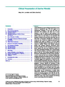
556 Normal blood flow
Truncus arteriosus
Blood return from body Aorta Blood pumped to the lungs
Pulmonary artery
Pulmonary veins P (blood returning ( from fro om lungs) o lu
LA Pulmonary valve
Aortic valve Ao
RA
Mitral Mit valve LV RV
Tricuspid Tric valve val
Blood return from body Dark blood with low oxygen content Pink blood oxygenated by the lungs
Fig. 46.1 Diagrammatic representation of normal circulation (right panel) and truncus arteriosus (left panel). Note that in truncus arteriosus, there is only one semilunar valve and this is referred to as the “truncal valve.” It can be tricuspid or quadricuspid (as is shown in the diagram). Abbreviations: RA right atria, LA left atria, RV right ventricle, LV left atrium (Reproduced or adapted from Driscoll D (2006) Fundamentals of pediatric cardiology. Lippincott Williams & Wilkins, with permission of the author and publisher)
common association between microdeletion 22q11.2 and truncus arteriosus especially if there is interruption of the aortic arch.
46.2
Pathologic Physiology
Cyanosis occurs because of a right-to-left shunt at the level of the ventricular septal defect and is dependent upon the volume of pulmonary blood flow. The relative volume of pulmonary and systemic blood flow depends on the relative resistance to flow into the pulmonary vascular bed and into the systemic vascular bed. The resistance to flow into the pulmonary vascular bed also will be effected by the presence, absence, and severity of pulmonary arte
Data Loading...