Enhanced patterns on intraoperative contrast-enhanced ultrasonography predict outcomes after curative liver resection in
- PDF / 1,184,848 Bytes
- 13 Pages / 595.276 x 790.866 pts Page_size
- 5 Downloads / 285 Views
ORIGINAL ARTICLE
Enhanced patterns on intraoperative contrast‑enhanced ultrasonography predict outcomes after curative liver resection in patients with hepatocellular carcinoma Ikuo Nakamura1 · Etsuro Hatano1 · Masaharu Tada1 · Yusuke Kawabata1 · Shinjiro Tamagawa1 · Ami Kurimoto1 · Hideaki Iwama1 · Kan Toriguchi1 · Hideaki Sueoka1 · Kenjiro Iida1 · Masahiro Yoshida2 · Takashi Nishimura3 · Hiroko Iijima3 Received: 25 March 2020 / Accepted: 4 September 2020 © Springer Nature Singapore Pte Ltd. 2020
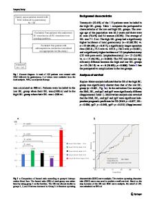
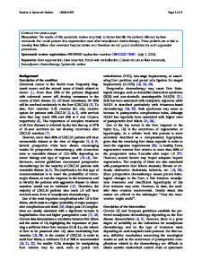
Abstract Purpose This study aimed to clarify what hepatocellular carcinoma (HCC) phenotype, as categorized by intraoperative contrast-enhanced ultrasonography (CEUS), showed a high risk of recurrence after hepatic resection. Methods Patients who underwent initial curative hepatectomy with intraoperative CEUS for a single HCC nodule were retrospectively assigned to three patterns of fine (FI), vascular (VA), and irregular (IR) according to the maximum intensity projection pattern based on intraoperative CEUS. Staining was performed for Ki-67, pyruvate kinase type M2 (PKM2), and vascular endothelial growth factor (VEGF) to assess the tumor proliferative activity, tumor glucose metabolism, and angiogenesis, respectively. Results Of 116 patients, 18, 50, and 48 were assigned to the FI, VA and IR patterns, respectively. IR patients demonstrated a significantly worse prognosis for both the recurrence-free survival (RFS) and overall survival (OS) (P = 0.0002, 0.0262, respectively) than did patients with other patterns. A multivariate analysis revealed an IR pattern in intraoperative CEUS to be an independent predictive factor for a poor RFS, and major hepatectomy and an IR pattern were independent predictive factors for a poor OS. An IR pattern was closely related to the tumor size (≥ 3.3 cm) and poor histological differentiation and showed a high Ki-67 index, low VEGF expression, and high PKM2 expression. Conclusion IR-pattern HCCs as classified by intraoperative CEUS may be associated with a higher risk of recurrence and worse outcomes in HCC patients after hepatic resection than other patterns. Keywords Hepatocellular carcinoma · Intraoperative contrast-enhanced ultrasonography · Liver resection
Introduction
Electronic supplementary material The online version of this article (https://doi.org/10.1007/s00595-020-02145-w) contains supplementary material, which is available to authorized users. * Etsuro Hatano shatano@hyo‑med.ac.jp 1
Department of Gastroenterological Surgery, Hyogo College of Medicine, Hyogo, Japan
2
Ultrasound Imaging Center, Hyogo College of Medicine, Hyogo, Japan
3
Division of Hepatobiliary and Pancreatic Disease, Department of Internal Medicine, Hyogo College of Medicine, Hyogo, Japan
Hepatocellular carcinoma (HCC) is one of the most common malignancies worldwide [1]. Various imaging modalities, including ultrasonography (US), computed tomography (CT), and magnetic resonance imaging (MRI), are critical for the diagnosis of HCC [2, 3]. US is the most frequently used imaging tool fo
Data Loading...