Induced Crystallization in CW Laser-Irradiated Sol-Gel Deposited Titania Films
- PDF / 1,164,431 Bytes
- 6 Pages / 414.72 x 648 pts Page_size
- 14 Downloads / 268 Views
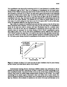
measurement of two Raman mode frequencies is sufficient to uniquely determine both temperature and residual stress as the amorphous film crystallizes. Since timeresolved Raman measurements can be carried out on very short time scales, this method is appropriate for following crystallization phenomena in amorphous films subjected to rapid step increases in temperature. Focused laser radiation offers the capability for producing rapid heating of films as well as the possibility for imprinting surface regions of variable refractive index. In work reported here, irradiation of amorphous sol-gel deposited films with a visible cw laser results in localized densification and crystallization at the irradiation sites. A critical laser fluence must be exceeded before crystallization can occur. Timeresolved Raman measurements acquired during sample irradiation are used to characterize surface temperature, the residual stress state of the film and how it evolves in time. Ongoing work involves applications of this technique for writing micrometer size images into refractory films deposited on silica or silicon substrates. Experimental Sol-gel titania films were prepared from highly acidic ethanol solutions of the titanium ethoxide precursor. 7 Homogeneous films were formed on silica or silicon(100) cleaned substrates at room temperature by spin casting at rates between 1200 and 1700 rev.min-1 for 60 s. Film thicknesses ranged from 200 to 1200 nm as determined from uv-vis-ir transmission measurements 8 and ellipsometry (632.8 nm and 700 incidence angle). Refractive indices averaged about 1.65 immediately after casting, and were found to increase somewhat with time at room temperature or during gentle heating. Isothermal heating of films to 1200C was achieved by means of a heat lamp located 25 cm above the coated substrates. Heating to higher temperatures involved the use of a muffle furnace or a resistively wound optical furnace for the in situ Raman measurements. 6 Surface morphology after heating was characterized by means of Transmission Electron Microscopy and Atomic Force Microscopy. 6,9 Laser heating involved imaging cw all-line emission from an Ar-ion laser onto the sol-gel coated substrate by means of an aberration-corrected 10x microscope objective having a small numerical aperture. Beam diameters were estimated at 10 gm. Under these conditions, laser fluences approaching several megawatts per cm 2 could be achieved. Film irradiations were performed at a number of fluences ranging from 0.1 to 5 MW.cm-2 and over times ranging from a few seconds to minutes. Spatially resolved Raman measurements of the laser irradiated regions were collected using an optical microscope interfaced to the entrance port of a Spex triple spectrometer. Spectra were excited using 100 mW of 514.5 nm argon ion laser radiation focused onto the sample by means of a 40x objective. Raman scattering was collected in a backscattering geometry. These conditions did not lead to densification or crystallization of the films during measurement. At significa
Data Loading...