Intermolecular Vibrations in Fullerene Systems
- PDF / 314,169 Bytes
- 6 Pages / 414.72 x 648 pts Page_size
- 31 Downloads / 413 Views
RESULTS AND DISCUSSION Shown in Fig. 1 is the intermolecular Raman spectrum of laser polymerized C6o. The intramolecular spectrum was found to be similar to that obtained on bulk powders [4] with an Ag( 2 ) mode at 1459cm-' and a lower frequency splitting of the Hg(1) mode of 12cm-'. However, the low frequency intermolecular scattering exhibits additional detail not previously observed in bulk material. In contrast to the major peak at - 115cm-1 noted previously, weaker features are observed in a broader band centered at - 90cm-' and a feature at - 140cm-'. Recent theoretical calculations for polymerized dimers of C60 have predicted Raman active modes at - 69cm-1 , 111cm- 1 and 123cm-' [6]. Previous evidence for the lowest frequency band was noted in temperature dependent studies of ultrathin polymerized C60 films deposited on Si0 2 [5]. These studies indicated changes in the relative intensity of the two lowest peaks with temperature. Variations in the relative intensity of the 90cm-' band with thickness have also been observed. The intermolecular spectra, as well as the splitting of the Hg(1) modes, support a dimer rather than an extended chain model for laser polymerized fullerenes. However, given the variations in the intensity of the lowest frequency peak, it is possible that other linked C60 cluster structures, such as linear and nonlinear trimers or larger units may occur. STM measurements of electron beam induced polymerized C60 exhibit larger unresolved clusters [9].
C80 photopolymer
U,,-
CO
" ' '
60
''
"...
....................
90
.
120
.
.
.
.
.
150
Roman Shift (cm-) Fig. 1 Intermolecular Raman spectrum of laser polymerized C60 (lower); the upper figure is background subtracted. In contrast to polymerized C60 modes, Fig. 2 shows the intermolecular modes in K3C60 obtained from Raman scattering on polycrystalline films and HREELS measurements on epitaxial films. The Raman and HREELS resolutions were 6cm' and 45cm-, respectively. The latter width is determined by a combination of spectrometer resolution and apparent surface roughness
494
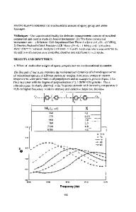
effects that vary with alkali-metal composition. The spectra shown were obtained after background subtraction of stray light and HREELS backgrounds discussed in more detail elsewhere [8]. As noted previously, the broad form of the Raman spectrum is due to disorder induced scattering. The spectrum is thus related to a Raman coupling parameter weighted density of states [1,7]. The HREELS spectrum peaks at considerably lower frequency than the Raman results. The latter has been related to the motion of K atoms at tetrahedral sites, while the former to K atoms at octahedral sites. The large difference in these frequencies is a
K3 C6 0
CO C"
V
30
60 90 120 Frequency (cm -1 )
150
Fig. 2 Comparison of HREELS (dashed) and Raman (solid) 300K spectra, background subtracted and normalized to a 1:2 area ratio. consequence of the considerably greater K - C distance for octahedral versus tetrahedral sites. The relative areas of 2:1 for the Raman and HREELS spec
Data Loading...