Mechanical modulation at the lamellar level in osteonal bone
- PDF / 408,299 Bytes
- 9 Pages / 585 x 783 pts Page_size
- 15 Downloads / 282 Views
P. Roschger Ludwig Boltzmann Institute of Osteology, A-1140 Vienna, Austria
H.D. Wagner Department of Materials and Interfaces, Weizmann Institute of Science, Rehovot 76100, Israel
P. Fratzl Department of Biomaterials, Max Planck Institute of Colloids and Interfaces, 14424 Potsdam, Germany (Received 20 December 2005; accepted 22 March 2006)
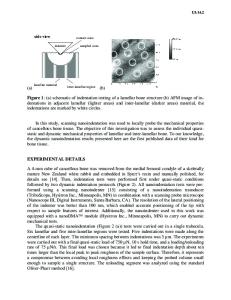
The secondary osteon is the fundamental building block of compact cortical bone at the tissue level. Light and scanning electron microscopy have shown that the osteon consists of a laminated cylindrical composite of mineralized collagen fibril lamellae ∼5–7 m thick. Using scanning nanoindentation and quantitative backscattered electron imaging on secondary osteons from the human femoral midshaft, we found that the indentation modulus shows a periodic variation between ∼24 GPa and ∼27 GPa within a single lamella. The average lamellar value remains nearly constant across the osteon and increases abruptly to more than 30 GPa at the interstitial bone interface. The local mineral content, determined from quantitative backscattered electron imaging at the indented locations, shows also a lamellar level modulation and is positively correlated with the indentation modulus at the same tissue position. We propose that such a mechanically and compositionally modulated structure may be an effective crack-stopping mechanism in bone.
I. INTRODUCTION
The compact cortical tissue in human bone consists of cylindrical secondary osteons embedded in a matrix of higher mineralized interstitial tissue.1 The mechanical properties of the osteon are crucial to maintaining the physiological and structural integrity of the compact bone tissue and, indeed, for bone as a whole. At the micrometer level, secondary osteons are multilayered cylindrical composites of mineralized fibrils arranged in lamellae about 3–5 m thick around a central Haversian system.2 Light microscopic investigation of the lamellar structure by Ascenzi and coworkers had suggested the existence of three osteon types: bright (fibers in the lamellae transverse to the osteon axis), dark (fibers parallel to the axis) and alternating (alternating fiber orientations),3,4 supporting the pioneering work of Gebhardt,5 indicating the existence of two perpendicularly oriented a)
Address all correspondence to this author. e-mail: [email protected] b) Present address: Department of Healthcare Devices & Instrumentation, Philips Research Laboratories, Prof.Holstaan 4 (WAG 01), 5656 AA Eindhoven, The Netherlands. DOI: 10.1557/JMR.2006.0234 J. Mater. Res., Vol. 21, No. 8, Aug 2006
http://journals.cambridge.org
Downloaded: 15 Mar 2015
orientations of fibers adjacent to each other. Higher resolution scanning2,6 and transmission7 electron microscopy on fracture surfaces of lamellar bone suggests strongly a “rotated plywood” structure where structurally orthotropic mineralized fibrils are rotated both with respect to the lamellar boundary as well as around their own fibril axis. Another model proposes that the lamellar structure arises du
Data Loading...