Muscle insulin resistance in type 1 diabetes with coronary artery disease
- PDF / 758,330 Bytes
- 10 Pages / 595.276 x 790.866 pts Page_size
- 96 Downloads / 300 Views
ARTICLE
Muscle insulin resistance in type 1 diabetes with coronary artery disease Katherine V. Williams 1,2 & Christina M. Shay 1,3 David E. Kelley 6 & Trevor J. Orchard 1
&
Julie C. Price 4,5 & Bret H. Goodpaster 6,7 & Carol A. Kelley 6 &
Received: 29 March 2020 / Accepted: 15 July 2020 # Springer-Verlag GmbH Germany, part of Springer Nature 2020
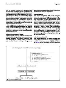
Abstract Aims/hypothesis The risk for coronary artery disease (CAD) is substantially increased in type 1 diabetes and it has been postulated that insulin resistance may contribute to this risk. The current study measured insulin resistance in type 1 diabetes with vs without CAD and with a focus upon skeletal muscle, to test the hypothesis that insulin resistance is more severe in participants who have type 1 diabetes and CAD. Additionally, in type 1 diabetes, we examined the hypothesis that insulin resistance is more severe in soleus (an oxidative type muscle) vs tibialis anterior (a more glycolytic type of muscle). Methods Insulin resistance was measured in participants with type 1 diabetes with (n = 9, CAD+) and without CAD (n = 10, CAD−) using euglycaemic insulin infusions combined with positron emission tomography (PET) imaging of [18F]fluorodeoxyglucose (FDG) uptake into soleus and tibialis anterior skeletal muscles. Coronary artery calcium (CAC) score was quantified by electron beam tomography. Results CAD+ participants with type 1 diabetes had a >100-fold higher CAC score than did CAD− participants with type 1 diabetes but groups did not differ in HbA1c or insulin dose. During clamp studies, CAD+ and CAD− groups had similar glucose disposal but were insulin resistant compared with historical non-diabetic participants (n = 13). FDG uptake by soleus muscle was similarly reduced, overall, in individuals with type 1 diabetes with or without CAD compared with non-diabetic individuals. However, FDG uptake by tibialis anterior muscle was not reduced in CAD− participants with type 1 diabetes while in CAD+ participants with type 1 diabetes it was 75% greater (p < 0.01). Across all participants with type 1 diabetes, FDG uptake by tibialis anterior muscle correlated positively with CAC severity. Conclusions/interpretation Our study confirms that systemic and skeletal muscle-specific insulin resistance is seen in type 1 diabetes but found that it does not appear to be more severe in the presence of CAD. There were, however, sharp differences between soleus and tibialis anterior muscles in type 1 diabetes: while insulin resistance was clearly manifest in soleus muscle, and was of equal severity in CAD+ and CAD− participants, tibialis anterior did not suggest insulin resistance in participants with type 1 diabetes, as FDG uptake by tibialis anterior correlated positively with CAC severity and was significantly increased in participants with type 1 diabetes and clinical CAD. Electronic supplementary material The online version of this article (https://doi.org/10.1007/s00125-020-05270-w) contains peer-reviewed but unedited supplementary material, which is available to authori
Data Loading...