Non-ECG-gated MDCTA of infracardiac total anomalous pulmonary venous connection in neonates and young infants
- PDF / 605,810 Bytes
- 5 Pages / 595.276 x 790.866 pts Page_size
- 93 Downloads / 301 Views
racardiac total anomalous pulmonary venous connection (TAPVC) is a rare congenital cardiac abnormality, in which the pulmonary veins have no connection with the left atrium and drain aberrantly into the tributaries of the inferior vena cava system. It often occurs with obstruction and other cardiac malformations, resulting in severe pulmonary edema that can hardly endure catheterization. Accurate assessment for morphological information of the pulmonary veins is crucial for the surgical strategy, but it still remains a challenge, especially in neonates and young infants. As noninvasive imaging modalities, both transthoracic echocardiography (TTE) and multidetector computed tomography angiography (MDCTA) have the potential to replace catheterization for TAPVC [1, 2, 3]; however, to our knowledge, there are only few reports on the infracardiac type of TAPVC because of the low incidence of this condition. Over the last 2 years, 9 infracardiac type patients were admitted to our cardiac center. This study aims to retrospectively compare the diagnostic value of non-ECG-gated MDCTA with TTE in the evaluation of infracardiac TAPVC.
Q. Yao · X. Hu · M. Pa · G. Huang Children’s Hospital of Fudan University, Shanghai
Non-ECG-gated MDCTA of infracardiac total anomalous pulmonary venous connection in neonates and young infants
went surgical correction. The research was approved by the ethics review committee and informed consent was given by the relatives of all patients.
Patients’ clinical data The age of the patients ranged from 2 days to 3 months (average 1.2±0.3 months). There were four girls and two boys, and
all babies were full-term. Among them, one had intrauterine growth retardation and one was an IVF baby. All patients presented with mild to moderate cyanosis and dyspnea, with an average heart rate (>140 beats/min) and respiratory rate (>50 breaths/min). The oxygen Qiong Yao and Xihong Hu contributed equally to this work.
Methods From March 2009 to October 2011, 9 consecutive patients were admitted to our cardiac center. After routine TTE suggesting infracardiac TAPVC, MDCTA was recommended for further detailed evaluation. All 9 patients under-
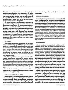
Fig. 1 8 Maximum intensity projection (a, b), and volume rendering (VR, c) reconstructed images show the whole vessels and the converging site (arrow) at the joint of the left and right portal vein. d Moderate interstitial edema in both fields of the lung without obvious venous obstruction Herz 5 · 2013
| 539
e-Herz: Original article pler, pulsed-wave Doppler, and continuous-wave Doppler.
Statistical analysis All images were assessed independently by two observers with more than 5 years’ experience of cardiac imaging, blinded to the surgical and the other imaging findings. An overall comparison between MDCTA and TTE was performed in three respects: the course of the drainage vein, the drainage site, and the presence of pulmonary vein stenosis including drainage vein hypogenesis, stenosis, or atresia [2]. Pulmonary vein stenosis was identified by a reduction of the ves
Data Loading...