On Mechanics of Connective Tissue: Assessing the Electrostatic Contribution to Corneal Stroma Elasticity
- PDF / 91,477 Bytes
- 6 Pages / 612 x 792 pts (letter) Page_size
- 74 Downloads / 313 Views
1239-VV03-07
On Mechanics of Connective Tissue: Assessing the Electrostatic Contribution to Corneal Stroma Elasticity Hamed Hatami-Marbini1 and Peter M. Pinsky Department of Mechanical Engineering, Stanford University, Stanford, CA 94305, USA
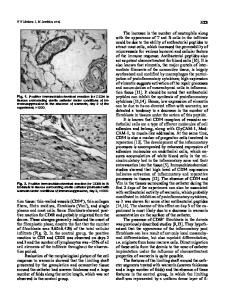
ABSTRACT The extracellular matrix plays a crucial role in defining the mechanical properties of connective tissues like cornea, heart, tendon, bone and cartilage among many others. The unique properties of these collagenous tissues arise because of both the hierarchal structure of collagens and the presence of negatively charged proteoglycans (PGs) which hold collagen fibers together. Here, in an effort to understand the mechanics of these structures, using the nonlinear Poison-Boltzmann (PB) equation, we study the electrostatic contribution to the elasticity of corneal stroma due to the presence of negatively charged PG glycosminoglycans (GAGs). Since collagens and GAGs have a regular hexagonal arrangement inside the corneal stroma, a triangular unit cell is chosen. The finite element method is used to solve the PB equation inside this domain and to obtain the electric potential and ionic distributions. Having the ion and potential distributions throughout the unit cell, the electrostatic free energy is computed and the tissue elasticity is calculated using the energy method. It is shown that as the ionic bath concentration increases; the electrostatic contribution to tissue elasticity is reduced. INTRODUCTION The mechanics of connective tissues (CTs) such as tendons, cartilage and cornea among others has been the subject of much recent research. The structure and behavior of these tissues are highly affected by both the mechanical and the electrostatic interactions of their constituents [1,2]. In this paper, we focus on the electrostatically-based compressive and in-plane shear properties of the corneal storma. Cornea consists of five parallel layers; namely, the epithelium, Bowman’s membrane, the stroma, Descemet’s membrane, and the endothelium. Corneal stroma occupies approximately 90% of the cornea thickness and is composed of collagen fibrils and PGs. The electrostatic and mechanical interaction of collagen fibrils and PGs defines the mechanical properties and function of connective tissues. Here, we quantitatively investigate the electrostatic origin of tissue elasticity through molecular-level modeling. Most of the previous studies on this subject have addressed the properties of cartilage [1,2,3,4]. Cartilage contains large aggregating proteoglycans (PGs) which are embedded in a cross-linked network of collagen fibers. In this tissue, both electrostatic and non-electrostatic forces balance the external compressive or shear load. While the electrostatic contribution arises from the interaction of negatively charged glycosaminoglycans (GAGs), i.e. the side chains of PGs, the non-electrostatic contribution to the tissue elasticity results from the inherent mechanical stiffness of collagen fibers and their entanglements with each other. The random fiber network model and
Data Loading...