Optical Control of Ligand-Gated Ion Channels
In the vibrant field of optogenetics, optics and genetic targeting are combined to commandeer cellular functions, such as the neuronal action potential, by optically stimulating light-sensitive ion channels expressed in the cell membrane. One broadly appl
- PDF / 417,050 Bytes
- 19 Pages / 504.57 x 720 pts Page_size
- 42 Downloads / 327 Views
1
Introduction Biology occurs over a wide range of time and length scales. To understand dynamic biological systems, we require tools for both the spatiotemporal observation and perturbation of cellular and molecular events. While the past years have seen rapid growth in optical (e.g., fluorescence-based) real-time reporters of cellular signals (1), the development of means to activate cells on short and small scales has lagged behind. With light being the premier choice for both readout and activation of cellular events, photosensitive molecules have recently enabled us to noninvasively control biological signals with high spatial and temporal resolution. This is achieved in optogenetics and optochemical genetics either by “repurposing” Nature’s light-sensing proteins from bacteria, algae, or plants or by engineering synthetic lightgated functionalities (2–4).
Nikita Gamper (ed.), Ion Channels: Methods and Protocols, Methods in Molecular Biology, vol. 998, DOI 10.1007/978-1-62703-351-0_32, © Springer Science+Business Media, LLC 2013
417
418
Stephanie Szobota et al.
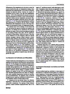
Fig. 1 Synthetic strategies for manipulating ligand-gated ion channels with light. (a) In the example of the ionotropic glutamate receptor GluK2, glutamate binding in the extracellular ligand binding domain triggers domain closure and gate opening. (b) Gating after ligand uncaging. (c) Gating after reversible conversion of a photochromic ligand to a binding-competent conformation. (d) Reversible light control by a PTL. (e) Chemical structure of MAG1, the prototypical maleimide-azobenzene-glutamate PTL, and 4-MG. (f, g) Crystal structure of the ligand binding domain of GluK2 in complex with 2S, 4R-4-methylglutamate (4-MG). 4-MG, residues surrounding the binding site suited for PTL attachment, and exit tunnel are highlighted as described in the text
Three synthetic strategies to light-control signals in cells or in vivo are commonly used: “caged” ligands, photochromic (“reversibly caged”) ligands, and photochromic tethered ligands (PTLs) (Fig. 1) (3). “Caged” ligands and photochromic ligands can be optically activated with subcellular resolution and within milliseconds either by releasing the ligand from the cage or by triggering a reversible conformational change in the photochrome. After photoactivation, these molecules act as free ligands on their specific protein targets (Fig. 1b, c). In contrast, PTLs are linear molecules that consist of a ligand moiety, a photochrome in the core of the molecule, and a reactive group that attaches to the protein (Fig. 1d, e). Site-directed attachment is achieved by genetically introducing a cysteine residue near the ligand binding site. After attachment, the agonist or antagonist located at the end of the
Remote Controlling Ion Channels
419
tether is presented to or retracted from the binding site by photoisomerization of the PTL core (Fig. 1d). Tethered ligands have the unique advantage that they specifically control a selected protein since the cysteine substitution is required for attachment and thus f
Data Loading...