Postmortem predation by a clowder of domestic cats
- PDF / 1,377,751 Bytes
- 4 Pages / 595.276 x 790.866 pts Page_size
- 58 Downloads / 388 Views
IMAGES IN FORENSICS
Postmortem predation by a clowder of domestic cats Roger W. Byard 1 Accepted: 11 August 2020 # Springer Science+Business Media, LLC, part of Springer Nature 2020
Abstract A 69-year-old man was found lying on the floor at his home address. According to the police report every room was filled with refuse and “thirty or so cats” were resident in the house. The body showed signs of extensive post mortem animal predation with opening of the chest and abdominal cavities, loss of soft tissues of the face, loss of soft tissues and organs of the neck, loss of the lungs and heart, and injuries to the liver, right kidney, stomach, transverse colon and cecum. The cause of death could not be determined from the autopsy given the absence of certain vital organs such as the heart and lungs, and the presence of early putrefaction. The case shows that considerable soft tissue, bone and organ loss may occur when a number of animals work in concert. The collective term for such a group of cats is a clowder. The extent of post-mortem damage from animal activity therefore relies not only on the species involved, but also the numbers of participating animals. Keywords Post-mortem predation . Animal activity . Cats . Clowder . Cause of death
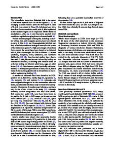
Case report A 69-year-old man was found lying on his back deceased at his home address on the lounge room floor (Fig. 1). According to the police report every room was filled with refuse and “thirty or so cats” were resident in the house. The body showed signs of extensive post mortem animal predation and a cat was actually present within the chest cavity. There was a past history of insulin-dependent diabetes mellitus and epilepsy. At autopsy the body was that of a decomposing adult white male showing extensive desiccated post mortem injuries due to animal predation with complete loss of skin and soft tissues of the neck, anterior chest and upper abdomen. The lesions were dried with no evidence of vital reaction or hemorrhage. The chest had been stripped back to the underlying sternum and ribs on the left side with a 25 × 30 mm defect in the left 2nd to 3rd intercostal space anterolaterally. There was also a
* Roger W. Byard [email protected] 1
Forensic Science SA and School of Medicine, The University of Adelaide, Level 2 Helen Mayo North Building, Frome Road, Adelaide, SA 5005, Australia
20 × 30 mm defect in the 6th to 7th intercostal space anterolaterally. On the right side the entire anterior rib cage had been removed exposing an empty chest cavity with absent heart and lungs (Fig. 2). The neck showed similar injuries with removal of the skin of the anterior neck and the right side, with loss of muscles and soft tissues through to the underlying cervical vertebrae. The esophagus, trachea, bronchi, larynx and thyroid gland were all absent and the transverse processes of the cervical vertebrae on the right side demonstrated animal bite marks. In the abdomen the intestines were exposed with skin and soft tissues stripped to the level of the umbilicus (Fig. 2). The live
Data Loading...