Primary Leiomyosarcoma of the Buccal Mucosa: Report of a Case and Review of the Literature
- PDF / 1,613,812 Bytes
- 5 Pages / 595.276 x 790.866 pts Page_size
- 87 Downloads / 326 Views
ORIGINAL PAPER
Primary Leiomyosarcoma of the Buccal Mucosa: Report of a Case and Review of the Literature Eugene M. Ko1 · Jonathan B. McHugh2 Received: 29 January 2018 / Accepted: 8 March 2018 © Springer Science+Business Media, LLC, part of Springer Nature 2018
Abstract This clinicopathologic study of primary oral leiomyosarcoma of the buccal mucosa involves a literature review of 15 cases with the addition of our report of a case. The demographic details, tumor size, treatment and outcome are documented for all the cases. In addition, this review examines the histologic features of leiomyosarcoma while noting that differentiation from other spindle cell tumors can be challenging, underscoring the necessity of an immunohistochemical work up for an accurate diagnosis. The unpredictability of the clinical behavior of these aggressive tumors requires, at the very least, wide local surgical excision and prolonged follow up. Keywords Leiomyosarcoma · Oral cavity · Head and neck
Introduction
Case Report
Leiomyosarcoma is a malignant tumor with smooth muscle differentiation and accounts for 5–10% of all soft-tissue sarcomas. It occurs most frequently in the retroperitoneum, and is exceedingly rare in the oral cavity [1, 2]. One explanation for the rarity of leiomyosarcomas in the oral cavity is attributed to the relative paucity of smooth muscles in that region [3–5]. Leiomyosarcomas that occur in the jawbones and oral tissues are thought to arise from the tunica media of blood vessels, erectores pilorum, circumvallate papillae, or from pluripotential mesenchymal cells [5]. Histopathologically, leiomyosarcomas typically present with interlacing fascicles of spindle cells, and so immunohistochemical discrimination of smooth muscle differentiation is often useful to help distinguish leiomyosarcomas from other tumors with similar light microscopic features. In part because of their rarity, leiomyosarcomas can be easily mistaken for other more common spindle cell lesions in the oral cavity [6].
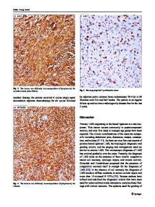
An 86-year old male presented to his oral surgeon with a swelling on his right posterior buccal mucosa, which had been unresponsive to antibiotics. An ulcerated area adjacent to tooth #1 was apparent. Subsequent incisional biopsy revealed an initial histopathologic diagnosis of a traumatic ulceration. However, a month later, the patient returned to the oral surgeon due to an increasing ulceration of the right buccal mucosa, and a re-biopsy was performed. No details regarding the size of the lesion nor radiographic images could be obtained from the oral surgeon. Microscopic examination revealed an infiltrative spindle cell neoplasm predominantly arranged in long fascicles and comprised of cells with abundant eosinophilic fibrillary cytoplasm and oval nuclei (see Figs. 1, 2, 3, 4, 5). Perinuclear vacuoles were present and there were scattered cells with marked cytologic atypia. No necrosis was present but mitotic figures were frequent (5–6 mitotic figures per 10 high-power fields). Immunohistochemical stains confirmed the morpholo
Data Loading...