Radiomics of high-resolution computed tomography for the differentiation between cholesteatoma and middle ear inflammati
- PDF / 593,117 Bytes
- 8 Pages / 595.276 x 790.866 pts Page_size
- 80 Downloads / 271 Views
HEAD AND NECK
Radiomics of high-resolution computed tomography for the differentiation between cholesteatoma and middle ear inflammation: effects of post-reconstruction methods in a dual-center study Christophe T. Arendt 1 & Doris Leithner 1,2 & Marius E. Mayerhoefer 2,3 & Peter Gibbs 2 & Christian Czerny 3 & Christoph Arnoldner 4 & Iris Burck 1 & Martin Leinung 5 & Yasemin Tanyildizi 6 & Lukas Lenga 1 & Simon S. Martin 1 & Thomas J. Vogl 1 & Ruediger E. Schernthaner 3 Received: 20 May 2020 / Revised: 7 November 2020 / Accepted: 25 November 2020 # The Author(s) 2020
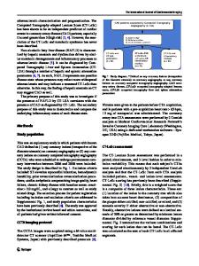
Abstract Objectives To evaluate the performance of radiomic features extracted from high-resolution computed tomography (HRCT) for the differentiation between cholesteatoma and middle ear inflammation (MEI), and to investigate the impact of postreconstruction harmonization and data resampling. Methods One hundred patients were included in this retrospective dual-center study: 48 with histology-proven cholesteatoma (center A: 23; center B: 25) and 52 with MEI (A: 27; B: 25). Radiomic features (co-occurrence and run-length matrix, absolute gradient, autoregressive model, Haar wavelet transform) were extracted from manually defined 2D-ROIs. The ten best features for lesion differentiation were selected using probability of error and average correlation coefficients. A multi-layer perceptron feed-forward artificial neural network (MLP-ANN) was used for radiomics-based classification, with histopathology serving as the reference standard (70% of cases for training, 30% for validation). The analysis was performed five times each on (a) unmodified data and on data that were (b) resampled to the same matrix size, and (c) corrected for acquisition protocol differences using ComBat harmonization. Results Using unmodified data, the MLP-ANN classification yielded an overall median area under the receiver operating characteristic curve (AUC) of 0.78 (0.72–0.84). Using original data from center A and resampled data from center B, an overall median AUC of 0.88 (0.82–0.99) was yielded, while using ComBat harmonized data, an overall median AUC of 0.89 (0.79–0.92) was revealed. Conclusion Radiomic features extracted from HRCT differentiate between cholesteatoma and MEI. When using multi-centric data obtained with differences in CT acquisition parameters, data resampling and ComBat post-reconstruction harmonization clearly improve radiomics-based lesion classification. Key Points • Unenhanced high-resolution CT coupled with radiomics analysis may be useful for the differentiation between cholesteatoma and middle ear inflammation. • Pooling of data extracted from inhomogeneous CT datasets does not appear meaningful without further post-processing.
Christophe Arendt and Doris Leithner contributed equally (shared first authorship). * Marius E. Mayerhoefer [email protected] 1
Department of Diagnostic and Interventional Radiology, University Hospital Frankfurt, Frankfurt am Main, Germany
2
Department of Radiology, Memorial Sloan Kettering Cancer Center,
Data Loading...